Review
. 2019 Dec 18;10(1):124.
doi: 10.1186/s13244-019-0817-4.

Intruding implements: a pictorial review of retained surgical foreign objects in neuroradiology

Affiliations
Review

Intruding implements: a pictorial review of retained surgical foreign objects in neuroradiology

Alessandra D'Amico et al. Insights Imaging. .

Abstract

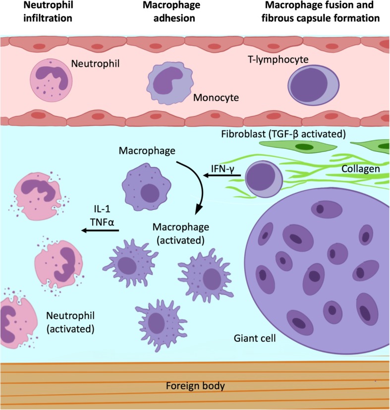
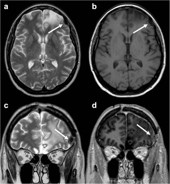
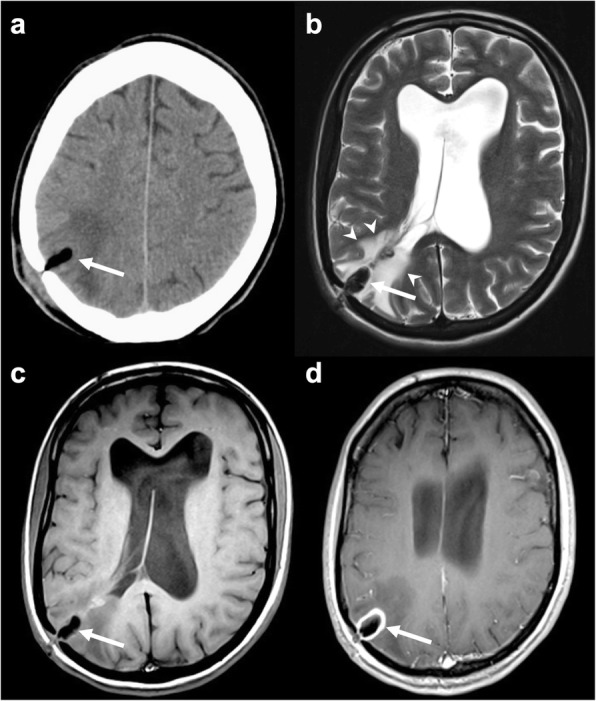
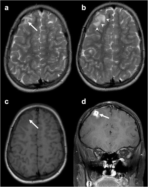
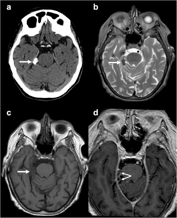
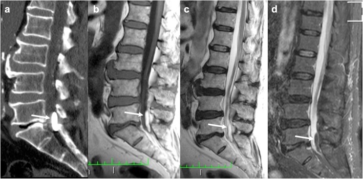
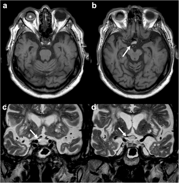
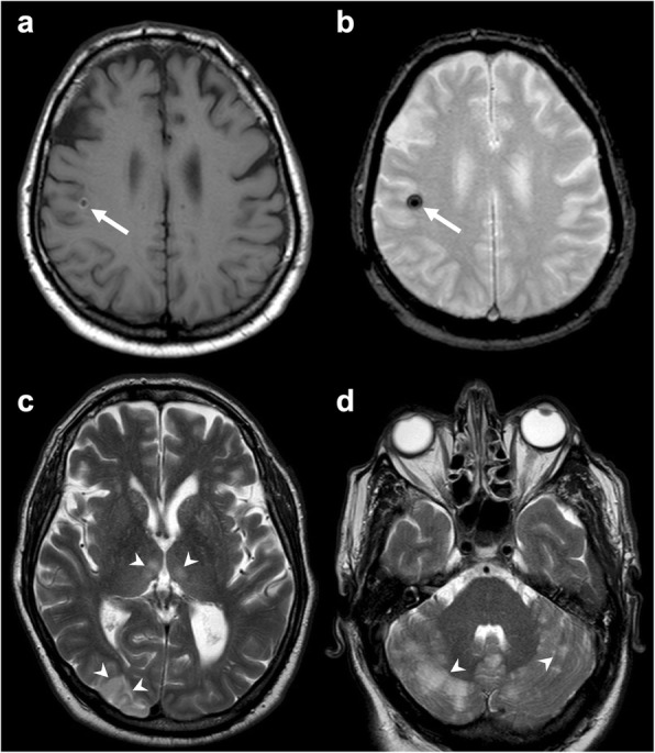
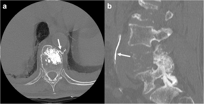
Intra-cranial and spinal foreign body reactions represent potential complications of medical procedures. Their diagnosis may be challenging as they frequently show an insidious clinical presentation and can mimic other life-threatening conditions. Their pathophysiological mechanism is represented by a local inflammatory response due to retained or migrated surgical elements. Cranial interventions may be responsible for the presence of retained foreign objects represented by surgical materials (such as sponges, bone wax, and Teflon). Spinal diagnostic and therapeutic procedures, including myelography, chordotomy, vertebroplasty, and device implantation, are another potential source of foreign bodies. These reactions can also follow material migration or embolization, for example in the case of Lipiodol, Teflon, and cement vertebroplasty. Imaging exams, especially CT and MRI, have a central role in the differential diagnosis of these conditions together with patient history. Neuroradiological findings are dependent on the type of material that has been left in or migrated from the surgical area. Knowledge of these entities is relevant for clinical practice as the correct identification of foreign bodies and related inflammatory reactions, material embolisms, or migrations can be difficult. This pictorial review reports neuroradiological semeiotics and differential diagnosis of foreign body-related imaging abnormalities in the brain and spine.

Keywords: Brain; Computed tomography; Foreign bodies; Magnetic resonance imaging; Spine.


Conflict of interest statement

The authors declare that they have no competing interests.

Figures

Fig. 1.
Fig. 1.
Foreign body granuloma pathophysiology. The foreign body response begins with rapid neutrophil infiltration and subsequent release of soluble factors involved in monocyte circulating monocyte recruitment. These last differentiate into macrophages, which are activated by T cell lymphocytes-produced interferon-γ. In turn, activated macrophages produce interleukin-1 family cytokines and tumor necrosis factor which determine further macrophage activation and their fusion into multinucleated giant cells. After months, transforming growth factor beta releasing recruits and activates fibroblasts with fibrous capsule development
Fig. 2
Fig. 2
Surgical sponge. Axial T2-weighted (a), unenhanced (b), and post-contrast (c) axial T1-weighted images showing an ovoidal mass (arrow) in the right superior temporal gyrus with low signal on both sequences without enhancement
Fig. 3.
Fig. 3.
Bone wax. Axial T2-weighted (a) and T1-weighted (b) and coronal T2-weighted (c) and post-contrast T1-weighted (d) images depicting burr hole bone wax, hypointense on both T1- and T2-weighted sequences (arrow in a, b, and c). Reactive enhancement is evident on post-contrast T1-weighted image (arrow in d)
Fig. 4
Fig. 4
Bone wax granuloma. Axial unenhanced CT (a), T2-weighted (b), unenhanced (c), and post-contrast (d) T1-weighted images presenting an ovoidal mass (arrow in a, b, and c) hypointense on all sequences and surrounded by vasogenic edema (arrowheads in b). After contrast administration, rim enhancement is detectable (arrow in d)
Fig. 5
Fig. 5
Suture material-induced granuloma. Axial T2-weighted (a, b) and unenhanced T1-weighted (c) images and coronal contrast-enhanced T1-weighted (d) showing a mass (arrow) in the right frontal superior gyrus with low signal on both T1- and T2-weighted sequences, surrounded by vasogenic edema (arrowheads), with rim enhancement. Some tubers are detectable in the axial images as the patient was affected by tuberous sclerosis
Fig. 6
Fig. 6
Teflon-induced calcified granuloma. Axial unenhanced CT (a), T2-weighted (b), unenhanced (c) and post-contrast (d) T1-weighted images depicting a calcified mass (arrow), with heterogeneous signal and rim enhancement
Fig. 7
Fig. 7
Ventricular catheter. Axial T2-weighted (a), FLAIR (b), unenhanced (c), and post-contrast (d) T1-weighted images presenting a left occipital access ventricular catheter (arrows) with mild peripheral gliosis (arrowheads in a and b) and subtle enhancement (arrowheads in d)
Fig. 8
Fig. 8
Lipiodol-induced adhesive arachnoiditis. Sagittal unenhanced CT (a), T1-weighted (b), T2-weighted (c), and STIR-weighted (d) images showing a mass in the spinal canal at the level of L5-S1 encasing the corresponding nerve roots. It appears hyperdense on CT and hyperintense on both T1- and T2-weighted sequences with signal suppression on STIR (arrows). The cauda nerve roots appear adherent to each other, consistent with the diagnosis of arachnoiditis
Fig. 9
Fig. 9
Silicon oil migration. Axial T1-weighted (a, b) and coronal T2-weighted (c, d) images demonstrating migration of silicon oil used to treat right retinal detachment (asterisk in a) through the right optic nerve sheath up to the right half of the optic chiasm (arrow in b, c, and d). The migrated material has heterogeneous signal also due to chemical shift artifact
Fig. 10
Fig. 10
Brain Teflon embolism. Axial T1-weighted (a), susceptibility-weighted (b), and T2-weighted (c, d) images depicting multiple ischemic lesions (arrowheads c, d) due to Teflon emboli (arrow in a and b) subsequent to aortic dissection surgery
Fig. 11
Fig. 11
Polymethylmethacrylate migration through veins. Axial (a) and sagittal (b) unenhanced CT presenting cement migration through an external anterior plexus vein (arrow in a) to the inferior vena cava (arrow in b)

References

    1. Eser O, Cosar M, Aslan A, Sahin O. Bone wax as a cause of foreign body reaction after lumbar disc surgery: a case report. Adv Ther. 2007;24:594–597. doi: 10.1007/BF02848783. - DOI - PubMed
    1. Onodera H, Furuya Y, Uchida M, et al. Intracranial foreign body granuloma caused by dural tenting suture. Br J Neurosurg. 2011;25:652–654. doi: 10.3109/02688697.2011.568641. - DOI - PubMed
    1. Smucker P, Bonnin JM, Pritz MB. Teflon granuloma with midbrain cysts after microvascular decompression of the trigeminal nerve. Acta Neurochir (Wien) 2007;149:537–539. doi: 10.1007/s00701-007-1136-y. - DOI - PubMed
    1. Anderson JM, Rodriguez A, Chang DT. Foreign body reaction to biomaterials. Semin Immunol. 2008;20:86–100. doi: 10.1016/j.smim.2007.11.004. - DOI - PMC - PubMed
    1. Grainger David W. All charged up about implanted biomaterials. Nature Biotechnology. 2013;31(6):507–509. doi: 10.1038/nbt.2600. - DOI - PubMed

LinkOut - more resources