. 2020 Jul;40(5):767-784.
doi: 10.1007/s10571-019-00771-8. Epub 2019 Dec 18.

Glioblastoma Stem Cell-Derived Exosomes Enhance Stemness and Tumorigenicity of Glioma Cells by Transferring Notch1 Protein

Affiliations

Glioblastoma Stem Cell-Derived Exosomes Enhance Stemness and Tumorigenicity of Glioma Cells by Transferring Notch1 Protein

Zhen Sun et al. Cell Mol Neurobiol. 2020 Jul.

Abstract

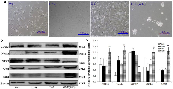
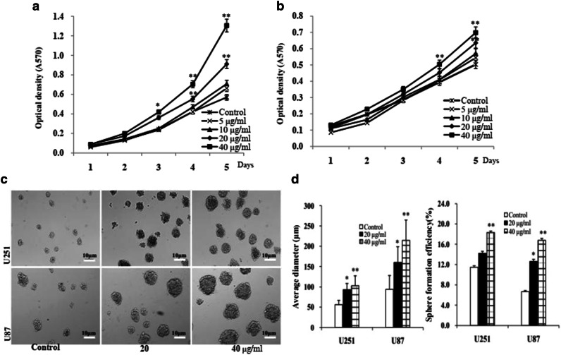
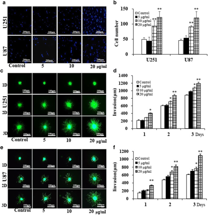
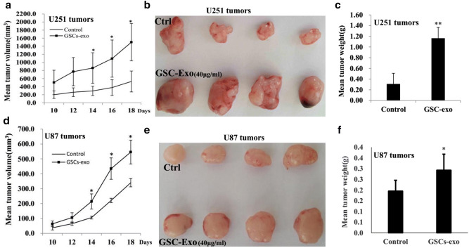
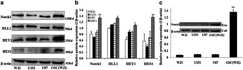
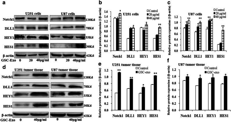
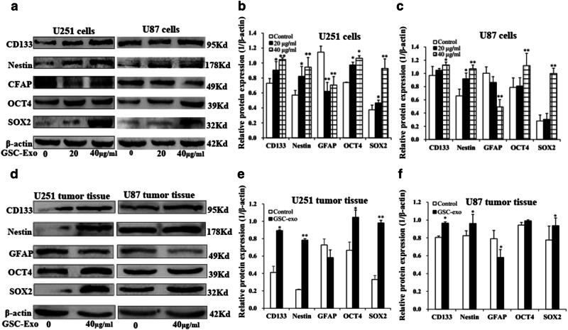
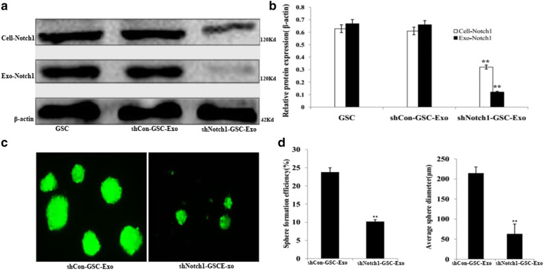
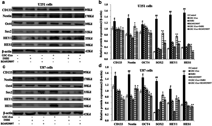
Exosomes contain plenty of bioactive information, playing an important role in intercellular communication by transfer their bioactive molecular contents to recipient cells. Glioblastoma stem cells (GSCs) and non-GSC glioma cells coexist in GBM microenvironment; GSC-released exosomes contain intracellular signaling molecules, which may affect the biological phenotypes of recipient cells. However, whether GSC exosomes could affect the biological phenotype of non-GSC glioma cells has not yet been defined. To explore whether GSC exosomes could reprogramme non-GSC glioma cells into GSCs and its possible mechanism involved, non-GSC glioma cells were treated with GSCs released exosomes; the potential mechanisms of action were studied with RNA interference, Notch inhibitors and Western blot analysis. The proliferation, neurosphere formation, invasive capacities, and tumorigenicity of non-GSC glioma cells were increased significantly after GSC exosome treatment; Notch1 signaling pathway was activated in GSCs; Notch1 protein was highly enriched in GSC exosomes; Notch1 signaling pathway and stemness-related protein expressions were increased in GSC exosome treated non-GSC glioma cells and these cell generated tumor tissues; Notch1 protein expression in GSCs and their exosomes, and the neurosphere formation of GSCs were decreased by Notch1 RNA interference; Notch1 signaling pathway protein and stemness protein expressions were decreased in GSC exosome treated non-GSC glioma cells by Notch1 RNA interference and Notch inhibitors. The findings in this study indicated that GSC exosomes act as information carriers, mediated non-GSC glioma cell dedifferentiation into GSCs by delivering Notch1 protein through Notch1 signaling activation, and enhanced stemness and tumorigenicity of non-GSC glioma cells.

Keywords: Dedifferentiation; Exosomes; Glioblastoma stem cells; Glioma cells; Notch1.


Conflict of interest statement

The authors declare that they have no conflicts of interest.

Figures

Fig. 1
Fig. 1
Morphological characteristic and stemness-related protein expressions of GSCs and non-GSC glioma cells. a Morphology of WJ1, U251, U87 cells, and GSCs. Glioma cells cultured in DMEM supplemented with 10% FBS grew as adherent monolayer, GSCs cultured in serum-free DMEM/F12 medium supplemented growth factors formed neurospheres. b Western blot analysis of stemness-related protein expressions in non-GSC glioma cells and GSCs. c The histogram shows that there was a significant difference of stemness-related protein expressions in non-GSC glioma cells and GSCs. (*p < 0.05, **p < 0.01)
Fig. 2
Fig. 2
Characterization of GSC exosomes and internalization in non-GSC glioma cells. a Transmission electron microscopy image of GSC exosomes. b size distribution of GSC exosomes. c Western blot analysis of CD63, TSG101, Alix, HSP70, and CytC of GSC exosomes and cells. d U251 and U87 non-GSC glioma cells were incubated with 20 μg/ml of CM-Dil-labeled GSC-exo for 6 h at 37 °C. Exosome internalization was observed by confocal microscopy
Fig. 3
Fig. 3
GSC exosomes promoted non-GSC glioma cell proliferation and neurosphere formation. a GSC exosomes promoted U251glioma cell proliferation. b GSC exosomes promoted U87 glioma cell proliferation. c GSC exosomes promoted U251and U87 glioma cell neurosphere formation. d The histogram shows the significant increase of neurosphere formation of U251 or U87 glioma cells treated with GSC exosomes. (*p < 0.05, **p < 0.01)
Fig. 4
Fig. 4
GSC exosomes promoted non-GSC glioma cell invasion. a U251 and U87 non-GSC glioma cells were treated with GSC exosomes for 3 days, and then, cell invasion was evaluated by transwell assay. b The histogram shows the significant increase of invasion numbers of U251 or U87 non-GSC glioma cells treated with GSC exosomes. c and e U251 and U87 non-GSC glioma cells were treated with GSC exosomes for 3 days, and then, cell invasion was measured by 3D-spheroid invasion assay. d and f The histogram shows the significant increase of invasion distance of U251 or U87 non-GSC glioma cells treated with GSC exosomes. (*p < 0.05, **p < 0.01)
Fig. 5
Fig. 5
GSC exosomes promoted non-GSC glioma cell tumorigenecity. 5 × 105 of the untreated and GSC exosomes treated U251 or U87 non-GSC glioma cells were inoculated into nude mice subcutaneously. ac Tumor growth of the untreated and GSC exosomes treated U251 non-GSC glioma cells. df Tumor growth of the untreated and GSC exosomes treated U87 non-GSC glioma cells. (*p < 0.05, **p < 0.01)
Fig. 6
Fig. 6
Notch1 signaling protein expressions in GSC and glioma cells and their exosomes. a Western blot analysis of Notch1 signaling protein expressions in non-GSC glioma cells and GSCs. b The histogram shows the significant increase of Notch1 signaling protein expressions in GSCs. c Notch1 protein was highly expressed in GSC exosomes, little was found in glioma cells derived exosomes. (*p < 0.05, **p < 0.01)
Fig. 7
Fig. 7
Notch1 signaling protein expressions in GSC exosomes treated non-GSC glioma cells and these cell generated tumor tissues. a Western blot analysis of Notch1 signaling protein expressions in GSC exosomes treated U251 and U87 non-GSC glioma cells. b and c The histograms show the significant increase of Notch1 signaling protein expressions in GSC exosomes treated U251 or U87 non-GSC glioma cells, respectively. d Notch1 signaling protein expressions in GSC exosomes treated U251 and U87 non-GSC glioma cell generated tumor tissues. The untreated and GSC exosomes treated U251 or U87 glioma cells were inoculated into nude mice, the generated tumor tissues were used for western blot analysis. e and f The histograms show the significant increase of Notch1 signaling protein expressions in GSC exosomes treated U251 and U87 non-GSC glioma cell generated tumor tissues. (*p < 0.05, **p < 0.01)
Fig. 8
Fig. 8
Stemness protein expressions in GSC exosomes treated non-GSC glioma cells and these cell generated tumor tissues. a Western blot analysis of stemness protein expressions in GSC exosomes treated U251 and U87 non-GSC glioma cells. b and c The histograms show the significant increase of stemness protein expressions in GSC exosomes treated U251 or U87 non-GSC glioma cells, respectively. d The stemness protein expressions in GSC exosomes treated U251 and U87 non-GSC glioma cell generated tumor tissues. The untreated and GSC exosomes treated U251 or U87 non-GSC glioma cells were inoculated into nude mice, the generated tumor tissues were used for western blot analysis. e and f The histograms show the significant increase of stemness protein expressions s in GSC exosomes treated U251 and U87 non-GSC glioma cell generated tumor tissues. (*p < 0.05, **p < 0.01)
Fig. 9
Fig. 9
shNotch1-GSC-exo internalization in GSCs. GSCs were incubated with 20 μg/ml of shNotch1-GSC-exo for 6 h at 37 °C, exosome internalization was observed by confocal microscopy
Fig. 10
Fig. 10
Notch1 RNA interference decreased Notch1 protein expression in GSCs and their exosomes and neurosphere formation of GSCs. a Western blot analysis of Notch1 protein expression in shNotch1-GSCs and their exosomes. b The histogram shows the significant increase of Notch1 protein expressions in shNotch1-GSCs and their exosomes. c Nerurosphere formation of shCon-GSC-Exo and shNotch1-GSC-Exo treated GSCs. d The histograms show the significant decrease of nerurosphere numbers and size of shNotch1-GSC-Exo treated GSCs. (*p < 0.05, **p < 0.01)
Fig. 11
Fig. 11
Notch1 RNA interference downregulates Notch1 signaling protein expressions in GSC exosomes treated non-GSC glioma cells. U251 and U87 non-GSC glioma cells treated with GSC exosomes (40 μg/ml) for 3 days, were further treated with shCon-GSC-Exo and shNotch1-GSC-Exo (40 μg/ml), respectively, and then Western blotting was performed. a Notch1 signaling protein expressions in GSC-Exo, shCon-GSC-Exo and shNotch1-GSC-Exo treated U251 non-GSC glioma cells. b The histogram shows the significant decrease of Notch1 signaling protein expressions in GSC-Exo treated U251 non-GSC glioma cells by shNotch1-GSC-Exo. c Notch1 signaling protein expressions in GSC-Exo, shCon-GSC-Exo and shNotch1-GSC-Exo treated U87 non-GSC glioma cells. d The histogram shows the significant decrease of Notch1 signaling protein expressions in GSC exosome treated U87 non-GSC glioma cells by shNotch1-GSC-Exo. (*p < 0.05, **p < 0.01)
Fig. 12
Fig. 12
Notch1 RNA interference downregulated stemness protein expressions in GSC exosomes treated non-GSC glioma cells. U251 and U87 non-GSC glioma cells treated with GSC exosomes (40 μg/ml) for 3 days, and further treated with shCon-GSC-Exo and shNotch1-GSC-Exo (40 μg/ml), respectively, and then Western blotting was performed. a Stemness protein expressions in shCon-GSC-Exo and shNotch1-GSC-Exo treated U251 non-GSC glioma cells. b The histogram shows the significant decrease of stemness protein expressions in GSC exosome treated U251 non-GSC glioma cells by shNotch1-GSC-Exo. c Stemness protein expressions in shCon-GSC-Exo and shNotch1-GSC-Exo treated U87 non-GSC glioma cells. d The histogram shows the significant decrease of stemness protein expressions in GSC exosome treated U87 non-GSC glioma cells by shNotch1-GSC-Exo. (*p < 0.05, **p < 0.01.)
Fig. 13
Fig. 13
Notch inhibitors decreased stemness and Notch1 target gene protein expressions in GSC exosomes treated non-GSC glioma cells. U251 and U87 non-GSC glioma cells were treated with GSC exosomes (40 μg/ml) for 3 days, and further treated with Notch inhibitor, IMR-1 or RO4929097, respectively, and then Western blotting was performed. a Stemness and Notch1 target gene protein expressions in GSC exosome treated U251 non-GSC glioma cells. b The histogram shows the significant decrease of stemness and Notch1 target gene protein expressions in GSC exosome treated U251 non-GSC glioma cells by Notch inhibitor, IMR-1 and RO4929097, *p < 0.05, **p < 0.01, compared with the control; #p < 0.05, ##p < 0.01, compared with GSC exosome treated U251 non-GSC glioma cells. c Stemness and Notch1 target gene protein expressions in GSC exosome treated U87 non-GSC glioma cells. d The histogram shows the significant decrease of stemness and Notch1 target gene protein expressions in GSC exosome treated U87 non-GSC glioma cells by Notch inhibitor, IMR-1 and RO4929097, *p < 0.05, **p < 0.01, compared with the control; #p < 0.05, ##p < 0.01, compared with GSC exosome treated U87 non-GSC glioma cells
Fig. 14
Fig. 14
schematic illustration demonstrates that GSC exosomes enhance the stemness and tumorigenicity of non-GSC glioma cells by transferring Notch1 protein through Notch1 signaling pathway activation

Similar articles

Cited by

References

    1. Andriani F, Bertolini G, Facchinetti F et al (2016) Conversion to stem-cell state in response to microenvironmental cues is regulated by balance between epithelial and mesenchymal features in lung cancer cells. Mol Oncol 10:253–271 - PMC - PubMed
    1. Astudillo L, Da Silva TG, Wang Z et al (2016) The small molecule IMR-1 inhibits the Notch transcriptional activation complex to suppress tumorigenesis. Cancer Res 76:3593–3603 - PMC - PubMed
    1. Auffinger B, Tobias AL, Han Y, Lee G, Guo D, Dey M et al (2014) Conversion of differentiated cancer cells into cancer stem-like cells in a glioblastoma model after primary chemotherapy. Cell Death Differ 21:1119–1131 - PMC - PubMed
    1. Bambury RM, Morris PG (2014) The search for novel therapeutic strategies in the treatment of recurrent glioblastoma multiforme. Expert Rev Anticancer Ther 14:955–964 - PubMed
    1. Bayin NS, Frenster JD, Sen R, Si S, Modrek AS, Galifianakis N et al (2017) Notch signaling regulates metabolic heterogeneity in glioblastoma stem cells. Oncotarget 8:4932–64953 - PMC - PubMed

MeSH terms

Substances