Review
. 2019 Dec 18;10(1):125-170.
doi: 10.1002/cphy.c190011.

Sex, Gender, and Sex Hormones in Pulmonary Hypertension and Right Ventricular Failure

Affiliations
Review

Sex, Gender, and Sex Hormones in Pulmonary Hypertension and Right Ventricular Failure

James Hester et al. Compr Physiol. .

Abstract

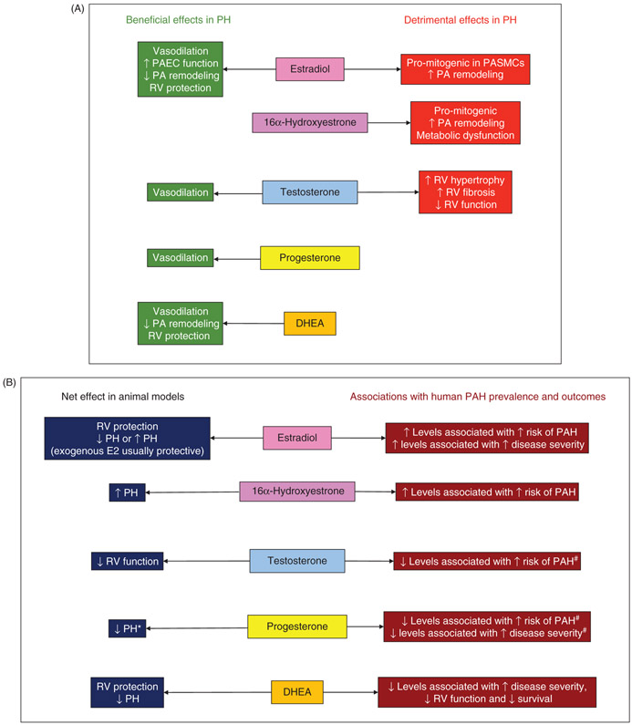
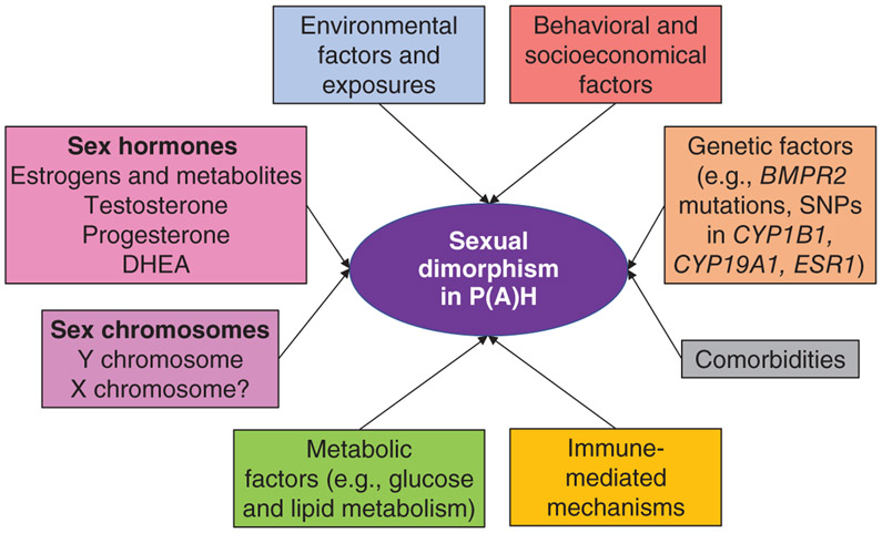
Pulmonary hypertension (PH) encompasses a syndrome of diseases that are characterized by elevated pulmonary artery pressure and pulmonary vascular remodeling and that frequently lead to right ventricular (RV) failure and death. Several types of PH exhibit sexually dimorphic features in disease penetrance, presentation, and progression. Most sexually dimorphic features in PH have been described in pulmonary arterial hypertension (PAH), a devastating and progressive pulmonary vasculopathy with a 3-year survival rate <60%. While patient registries show that women are more susceptible to development of PAH, female PAH patients display better RV function and increased survival compared to their male counterparts, a phenomenon referred to as the "estrogen paradox" or "estrogen puzzle" of PAH. Recent advances in the field have demonstrated that multiple sex hormones, receptors, and metabolites play a role in the estrogen puzzle and that the effects of hormone signaling may be time and compartment specific. While the underlying physiological mechanisms are complex, unraveling the estrogen puzzle may reveal novel therapeutic strategies to treat and reverse the effects of PAH/PH. In this article, we (i) review PH classification and pathophysiology; (ii) discuss sex/gender differences observed in patients and animal models; (iii) review sex hormone synthesis and metabolism; (iv) review in detail the scientific literature of sex hormone signaling in PAH/PH, particularly estrogen-, testosterone-, progesterone-, and dehydroepiandrosterone (DHEA)-mediated effects in the pulmonary vasculature and RV; (v) discuss hormone-independent variables contributing to sexually dimorphic disease presentation; and (vi) identify knowledge gaps and pathways forward. © 2020 American Physiological Society. Compr Physiol 10:125-170, 2020.


Figures

Figure 1
Figure 1. Current classification of pulmonary hypertension (PH) and subtypes with evidence for sexually dimorphic features.
PH classification from 6th World Symposium (Nice, 2018) according to Simonneau et al. (351). In addition to the data presented here, one study in a large cohort of veterans with all types of PH (predominantly Group 2 and 3 PH; n = 15,464 patients) demonstrated that women with PH exhibit higher pulmonary vascular resistance and pulmonary artery pulse pressure, yet lower RAP as well as 18% greater survival compared to men with PH. *These analyses predominantly included patients with idiopathic PAH and also patients with heritable PAH and drug- and toxin-associated PAH (no subgroup analyses performed). #Attenuated hypoxia-induced PH in women not consistently found across studies. BMPR2, gene encoding bone morphogenic protein receptor 2; CYP1B1, gene encoding cytochrome P450 1B1; CYP19A1, gene encoding aromatase; ESR1, gene encoding estrogen receptor α; HFpEF, heart failure with preserved ejection fraction; HIV, human immunodeficiency virus; HT, hormone therapy; LVEF, left ventricular ejection fraction; PCH, pulmonary capillary hemangiomatosis; PVOD, pulmonary veno-occlusive disease; PVR, pulmonary vascular resistance; RV, right ventricle; SNP, single-nucleotide polymorphism; SSc, systemic sclerosis.
Figure 2
Figure 2. Pathophysiology of PAH.
(A) Arterial cross section illustrating PAH pathology in the pulmonary arteries. Proliferation of endothelial cells (ECs), smooth muscle cells (SMCs), and fibroblasts leads to vascular remodeling with eventual occlusion of diseased vessels. Neoangiogenesis driven by apoptosis-resistant proliferative ECs, SMCs, and other resident PA and recruited cells promotes formation of plexiform vascular lesions, which are the hallmark of PAH. Plexiform lesions may be seen within pulmonary vessels as well as extending into the adventitial tissue (not shown). Infiltration of PH vascular lesions by immune cells and bone marrow-derived cells drives a pro-inflammatory and pro-proliferative state in the tissue. (B) Transverse section of the heart. High pulmonary vascular resistance in PAH produces increased afterload on the RV, resulting in adaptation and RV failure in PAH. RV hypertrophy may be adaptive and compensatory to overcome PVR and maintain cardiac output (not shown). On the other hand, RV hypertrophy may be maladaptive, marked by vessel rarefaction, metabolic dysfunction, inflammation, cell death, fibrosis, and increased RV dilatation. Maladaptive RV remodeling is associated with RV ischemia and decreased RV ejection fraction and cardiac output, resulting in RV failure.
Figure 3
Figure 3. Sex hormone synthesis and estrogen metabolism.
Steroidogenic enzymes represented here are present in the lung, heart, and/or vascular tissue (148, 197, 314, 317, 480). Abundance of boxed enzymes is altered in PH/PAH (12, 326, 340, 457, 462-464). Compounds in red have been targeted in clinical trials of PAH therapies (193). 2-Hydroxyestradiol and 2-methoxyestradiol exert ER-independent antiproliferative, anti-inflammatory effects that appear to mitigate vascular remodeling in animal models of PAH (22, 82, 421). Conversely, the estrogen metabolites 4-hydroxyestradiol, 4-methoxyestradiol, and 16α-hydroxyestrone signal via estrogen receptors and promote a mitogenic, inflammatory, and antiapoptotic phenotype that exacerbates PH/PAH (54, 463). *Multiple CYP enzymes are capable of catalyzing estrogen hydroxylation; CYP1A1 and CYP1B1 appear to be the most relevant isoforms in PAH pathology. Factors including diet (240, 275), hypoxia (107), inflammation (11), genetics (12, 146), and drug exposure (71) may alter estrogen metabolism. Effects of enzymes and metabolites depicted here may be cell-, tissue-, and/or organ specific.
Figure 4
Figure 4. Overview of major biological effects of the most abundant sex hormones (A) and their net effects in animal studies as well as reported associations with PAH risk and outcomes in human studies (B).
Note that effects of specific sex hormones on outcomes in human studies may be limited to men or pre- or postmenopausal women only. Human studies measured DHEA-S (dehydroepiandrosterone sulfate). *Data based on study only. #Inconsistent associations across studies. DHEA, dehydroepiandrosterone; PA, pulmonary artery; PAEC, pulmonary artery endothelial cell; PAH, pulmonary arterial hypertension (human studies); PASMCs, pulmonary artery smooth muscle cells; PH, pulmonary hypertension; RV, right ventricle.
Figure 5
Figure 5. Simplified overview of factors contributing to sex/gender differences and sexual dimorphism in PAH and PH.
Note that significant cross talk exists between the factors and mediators listed in this figure. BMPR2, gene encoding bone morphogenic protein receptor 2; CYP1B1, gene encoding cytochrome P450 1B1; CYP19A1, gene encoding aromatase; DHEA, dehydroepiandrosterone; ESR1, gene encoding estrogen receptor α; SNP, single-nucleotide polymorphism.

References

    1. Abenhaim L, Moride Y, Brenot F, Rich S, Benichou J, Kurz X, Higenbottam T, Oakley C, Wouters E, Aubier M, Simonneau G, Begaud B. Appetite-suppressant drugs and the risk of primary pulmonary hypertension. International Primary Pulmonary Hypertension Study Group. N Engl J Med 335: 609–616, 1996. - PubMed
    1. Achar S, Rostamian A, Narayan SM. Cardiac and metabolic effects of anabolic-androgenic steroid abuse on lipids, blood pressure, left ven-tricular dimensions, and rhythm. Am J Cardiol 106 (6): 893–901, 2010. - PMC - PubMed
    1. Ahn BH, Park HK, Cho HG, Lee HA, Lee YM, Yang EK, Lee WJ. Estrogen and enalapril attenuate the development of right ventricular hypertrophy induced by monocrotaline in ovariectomized rats. J Korean MedSci 18: 641–648, 2003. - PMC - PubMed
    1. Albrecht ED, Pepe GJ. Placental steroid hormone biosynthesis in primate pregnancy. Endocr Rev 11 (1): 124–150, 1990. - PubMed
    1. Alencar AK, Montes GC, Montagnoli T, Silva AMS, Martinez ST, Fraga AG, Wang H, Groban L, Sudo RT, Zapata-Sudo G. Activation of GPER ameliorates experimental pulmonary hypertension in male rats. Eur J Pharm Sci 97: 208–217, 2017. - PMC - PubMed

MeSH terms

Substances