Review
. 2020 Jan;21(1):76-93.
doi: 10.1111/tra.12715.

Life in the lumen: The multivesicular endosome

Affiliations
Review

Life in the lumen: The multivesicular endosome

Jean Gruenberg. Traffic. 2020 Jan.

Abstract

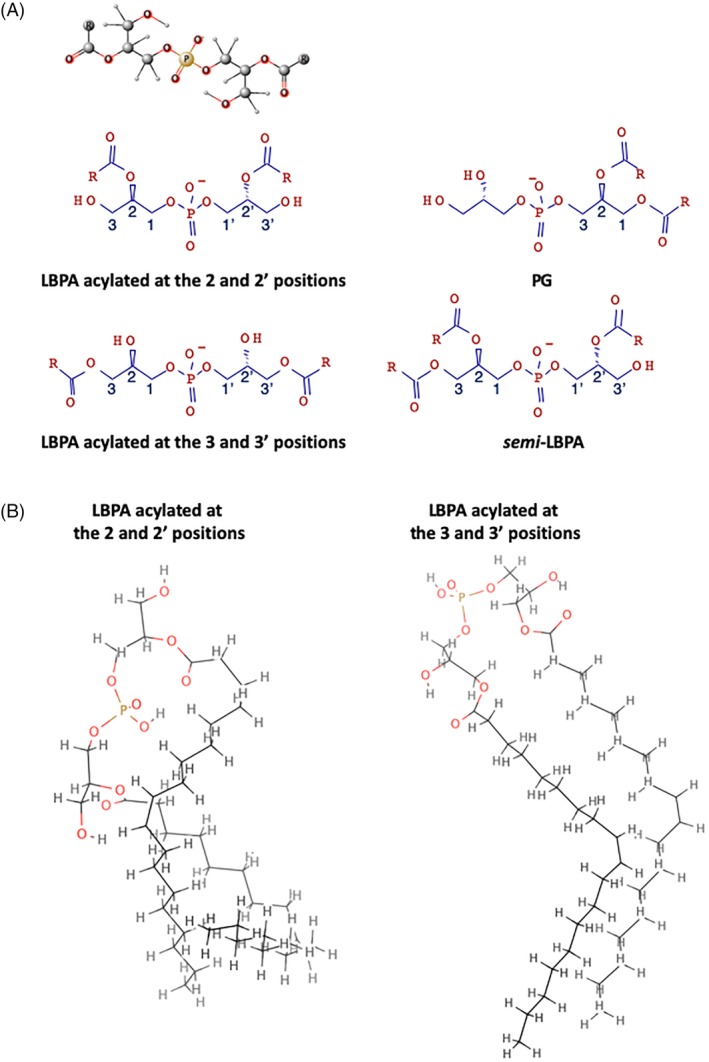
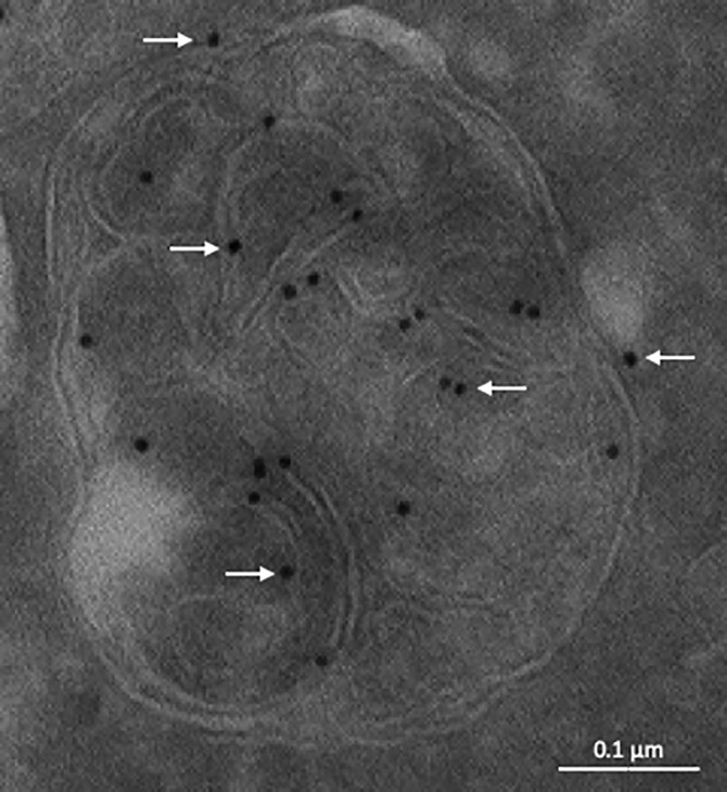
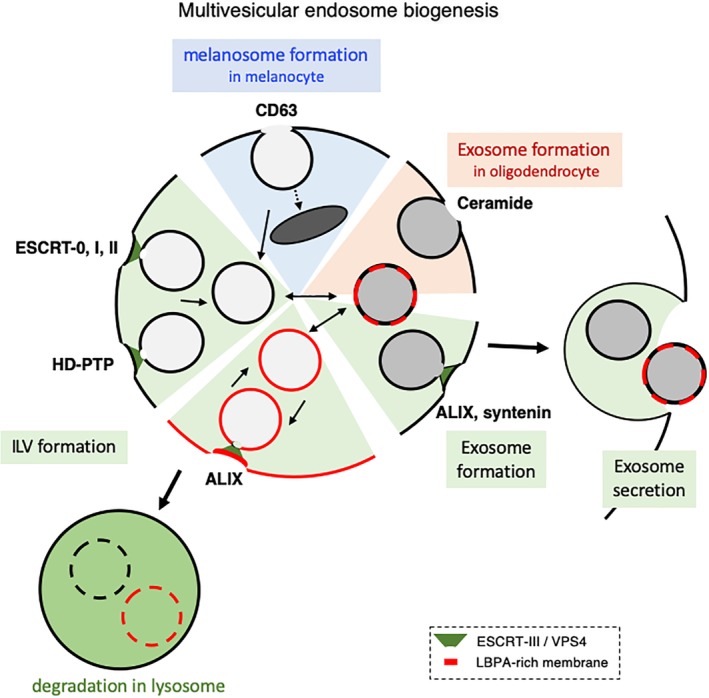
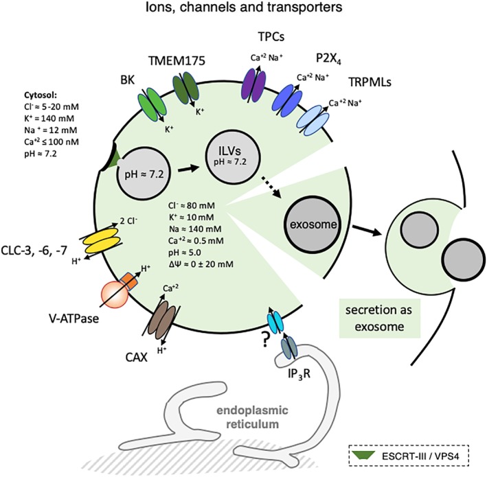
The late endosomes/endo-lysosomes of vertebrates contain an atypical phospholipid, lysobisphosphatidic acid (LBPA) (also termed bis[monoacylglycero]phosphate [BMP]), which is not detected elsewhere in the cell. LBPA is abundant in the membrane system present in the lumen of this compartment, including intralumenal vesicles (ILVs). In this review, the current knowledge on LBPA and LBPA-containing membranes will be summarized, and their role in the control of endosomal cholesterol will be outlined. Some speculations will also be made on how this system may be overwhelmed in the cholesterol storage disorder Niemann-Pick C. Then, the roles of intralumenal membranes in endo-lysosomal dynamics and functions will be discussed in broader terms. Likewise, the mechanisms that drive the biogenesis of intralumenal membranes, including ESCRTs, will also be discussed, as well as their diverse composition and fate, including degradation in lysosomes and secretion as exosomes. This review will also discuss how intralumenal membranes are hijacked by pathogenic agents during intoxication and infection, and what is the biochemical composition and function of the intra-endosomal lumenal milieu. Finally, this review will allude to the size limitations imposed on intralumenal vesicle functions and speculate on the possible role of LBPA as calcium chelator in the acidic calcium stores of endo-lysosomes.

Keywords: ALIX; ESCRTs; Niemann-pick C; anthrax; bis(monoacylglycero)phosphate BMP; calcium store; cholesterol; enveloped virus; exosome; intralumenal vesicle ILV; lipidomics; lysobisphosphatidic acid; lysosome; lysosome storage disease; multivesicular endosome; pathogen; penetration; toxin.


Figures

Figure 1
Figure 1
Outline of the endocytic pathway. Organization of the endosomal pathway in mammalian cells, but not in yeast or plant cells.2 Endocytosed components are delivered to a common early endosome, from where some proteins and lipids are recycled back to the plasma membrane, or routed by retrograde transport to the trans‐Golgi network. Molecules destined for late endosomes are sorted into ILVs forming on early endosomal membranes, giving rise to multivesicular endosomes. These detach (or mature) from early endosomes and transports cargoes toward late endosomes and lysosomes. Eventually, some ILVs are delivered to lysosomes where they are degraded together with their protein cargo. Late endosomes and lysosomes exchange membrane components and solutes, forming a transient hybrid endo‐lysosome, which is then re‐converted into secondary lysosomes, where hydrolases are stored. Endosomes and lysosomes can also undergo fusion with the plasma membrane as secretory endo‐lysosomes, and ILVs can also be released extracellularly as exosomes. The endosomal pathway also serves as an input or output for other membrane trafficking pathways, as indicated. In particular, endosomes and lysosomes also function at a crossroad with the autophagy pathway, and engage in physical contacts via membrane contact sites with other organelles, including the endoplasmic reticulum
Figure 2
Figure 2
LBPA and isoforms. A, LBPA vs PG and LBPA isoforms. The ball‐and‐stick model of LBPA acylated at the 2 and 2′ positions is shown on top of the figure, above the schematic representations of the same isoform, as well as LBPA acylated at the 3 and 3′ positions, PG and semi‐LBPA. B, LBPA acylated at the 2 and 2′ positions vs LBPA acylated at the 3 and 3′ positions. The outlines show the atomistic description by molecular dynamics at the quantum mechanical level of two of the lowest energy conformers for both 2,2’‐LBPA and 3,3’‐LBPA83
Figure 3
Figure 3
Distribution of LBPA in late endosomes illustrated by immunogold labeling of cryosections. The electron micrograph shows a late endosome of HeLa cells labeled with the anti‐LBPA monoclonal antibody 6C4, followed by 10 nm protein A‐gold (arrows). Bar: 0.1 μm. [Courtesy of Robert G. Parton, Brisbane, Australia]
Figure 4
Figure 4
Multivesicular endosome biogenesis. The figure outlines the proposed mechanisms driving the formation of ILVs and exosomes in most cell types (green), exosomes in oligodendrocytes (brown) and melanosomes in melanocytes (blue). In most cell types, sorting into ILVs is mediated by ESCRT‐0, ‐I and ‐II, HD‐PTP or ALIX, as is presumably the nucleation of ESCRT‐III filaments, which drive the membrane deformation process. However, ILVs may also be formed in a CD63‐dependent and ESCRT‐independent manner—a process presumably akin to the biogenesis of melanosomes in melanocytes. ILVs formed in early endosomes presumably lack LBPA, because the lipid is only found in late endosomes. The biogenesis of exosomes may require ALIX and ESCRTs, as well as syntenin presumably, but not in oligodendrocytes where the process seems to depend on ceramides and to be ALIX‐ and ESCRT‐independent. Once formed, ILVs and exosomes follow different pathways. ILVs can be targeted to lysosomes for degradation, or undergo back‐fusion with the limiting membrane. Exosomes by contrast are secreted upon endosome fusion with the plasma membrane. The relationship between ILVs and exosomes are not clear. Neither are the mechanisms that discriminate their selective fates. The factors that have been reported to control each process are indicated. Membranes shown in the black color imply that it is not known whether the corresponding processes involve LBPA‐containing membranes
Figure 5
Figure 5
Viruses, toxin and ILV‐membrane dynamics. The left side of the figure (penetration) outlines the pathways used by some endocytosed pathogenic agents that enter the host‐cell cytoplasm through endosomes, in a process that depends on proteins/lipids involved in ILV membrane dynamics. VSV, Lassa virus, LCMV, and Flaviviruses may penetrate cells in a two‐step process. First, the viral enveloped undergoes fusion with the ILV membrane (eg, in early endosomes) so that the capsid be delivered into the protected environment of the ILV lumen. Then, the capsid is released into the host‐cell cytoplasm upon fusion of the ILV membrane with the late endosome limiting membrane (so‐called back‐fusion). Similarly, the anthrax toxin is first translocated across the ILV membrane and then delivered to the cytoplasm upon ILV back‐fusion. Other endocytosed viruses may penetrate cells upon direct fusion of the viral envelope with the late endosome membrane.206 The lower part of the figure outlines the role of ESCRT‐III and other ESCRT sub‐units in repairing damage to vacuoles containing the indicated bacteria. The right side of the figure outlines the inclusion of some viruses and viral particles into exosomes (enclosure) in a process that depends on ESCRT components, and their release as exosomes. The endocytosed anthrax toxin can also be released as exosomes, rather than being delivered to the cytoplasm of the target cell. Membranes shown in the black color imply that it is not known whether the corresponding processes involve LBPA‐containing membranes. CCHFV, Crimean‐Congo hemorrhagic fever virus; HAV, hepatitis A virus; HCV, hepatitis C virus; HPV, human papillomaviruses; IAV, influenza A virus; LCMV, lymphocytic choriomeningitis; VSV, vesicular stomatitis virus; M marinum, Mycobacterium marinum (in Dictyostelium discoideum cells); M tuberculosis, Mycobacterium tuberculosis); C burnetii, Coxiella burnetii
Figure 6
Figure 6
Ions, channels and transporters. The figure outlines the major ion channels and transporters present in endo‐lysosome, as well as the estimated ion concentration in the lumen of endo‐lysosomes and in the cytoplasm. The intralumenal concentration of Cl was estimated using a DNA‐based, fluorescent chloride reporter271 and see also.272 The lumenal concentration of Na is estimated to be around 140‐150 mM.245 Li and collaborators recently proposed that ΔΨ of resting lysosomes is around 0 (±20 mV).235 Essentially nothing is known about the ionic situation within ILVs or exosomes, except for the observation that ILVs remain neutral until at least 20 minutes after formation.266 At ER‐lysosome membrane contact sites, the ER may sequester lysosomal Ca2+,273 and ER Ca2+ may refill lysosomal Ca2+ stores.274 Ca2+ is released from ER stores via Ins(1,4,5)P3 receptor (IP3R) and calcium refilling of the endosomes may be driven by the proton gradient via a vertebrate Ca2+/H+ exchanger (CAX),275 or depend directly on the ER in a pH‐independent fashion.276 Membranes shown in the black color imply that it is not known whether the corresponding processes involve LBPA‐containing membranes. V‐ATPase: the vacuolar ATPase240; CLC‐3, ‐6, ‐7: the 2Cl/H+‐exchangers CLC‐3, ‐6, ‐7 (chloride channels) that distribute in endo‐lysosomes238; CAX, a putative endo‐lysosomal Ca2+/H+ exchanger involved in Ca2* uptake into endo‐lysosomes275; P2X4, purinergic P2X receptor subtype 4; TPC, two‐pore channels; TRPMLs, transient receptor potential channels; BK, big conductance Ca2+‐activated potassium channel274; TMEM175: K+‐selective channel235

References

    1. Huotari J, Helenius A. Endosome maturation. EMBO J. 2011;30(17):3481‐3500. - PMC - PubMed
    1. Scott CC, Vacca F, Gruenberg J. Endosome maturation, transport and functions. Semin Cell Dev Biol. 2014;31:2‐10. - PubMed
    1. Simonetti B, Cullen PJ. Endosomal sorting: architecture of the Retromer coat. Curr Biol. 2018;28(23):R1350‐R1352. - PubMed
    1. Hanson PI, Cashikar A. Multivesicular body morphogenesis. Annu Rev Cell Dev Biol. 2012;28:337‐362. - PubMed
    1. Luzio JP, Pryor PR, Bright NA. Lysosomes: fusion and function. Nat Rev Mol Cell Biol. 2007;8(8):622‐632. - PubMed

Publication types

LinkOut - more resources