Review
. 2019 Dec;20(12):1597-1615.
doi: 10.3348/kjr.2018.0931.

Rapid Imaging: Recent Advances in Abdominal MRI for Reducing Acquisition Time and Its Clinical Applications

Affiliations
Review

Rapid Imaging: Recent Advances in Abdominal MRI for Reducing Acquisition Time and Its Clinical Applications

Jeong Hee Yoon et al. Korean J Radiol. 2019 Dec.

Abstract

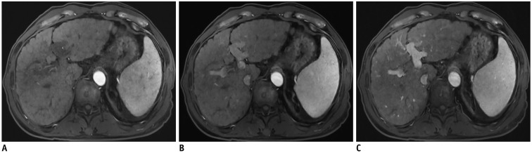
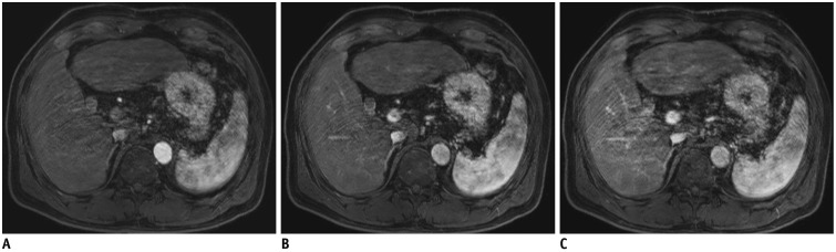
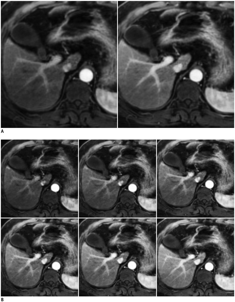
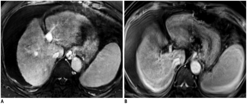
Magnetic resonance imaging (MRI) plays an important role in abdominal imaging. The high contrast resolution offered by MRI provides better lesion detection and its capacity to provide multiparametric images facilitates lesion characterization more effectively than computed tomography. However, the relatively long acquisition time of MRI often detrimentally affects the image quality and limits its accessibility. Recent developments have addressed these drawbacks. Specifically, multiphasic acquisition of contrast-enhanced MRI, free-breathing dynamic MRI using compressed sensing technique, simultaneous multi-slice acquisition for diffusion-weighted imaging, and breath-hold three-dimensional magnetic resonance cholangiopancreatography are recent notable advances in this field. This review explores the aforementioned state-of-the-art techniques by focusing on their clinical applications and potential benefits, as well as their likely future direction.

Keywords: Compressed sensing; Diffusion weighted imaging; Free breathing; GRASE; GRASP; MRCP; MRI; Simultaneous multi-slice; Sparse.


Conflict of interest statement

Two authors are employees of Siemens Healthcare (Nickel MD) and Philips Healthcare (Peeters JM).

Figures

Fig. 1
Fig. 1. Different k-space sampling schemes.
Cartesian acquisition with fully sampled k-space (A), uniformly undersampled with acceleration factor of 2 (B), or non-Cartesian acquisition with skipping spokes in radial acquisition (C). Solid and dashed lines refer to acquired and skipped k-space data, respectively. Kx = frequency encoding direction, Ky = phase encoding direction
Fig. 2
Fig. 2. Graphical representation of principles of parallel imaging and compressed sensing.
In parallel imaging (A), uniform subsampling gives typical aliasing artifacts. Parallel imaging reconstruction makes it possible to achieve alias-free image. For SENSE, parallel imaging reconstruction is done in image space, whereas de-aliasing is already done before FFT in generalized autocalibrating partially parallel acquisitions (GRAPPA). In compressed sensing (B), variable-density, pseudo-random subsampling produces incoherent noise-like aliasing artifact after FFT. Sparsity (in this case, wavelet) transform allows setting of sparsity constraints. Image is obtained after IWT into image domain. IFFT back to k-space allows data consistency checking. After several iterations, final image is delivered with optimal balance between data consistency and sparsity constraints. FFT = Fourier transform, IFFT = inverse Fourier transform, IWT = inverse transform, PI = parallel imaging, SENSE = sensitivity encoding, WT = wavelet transform
Fig. 3
Fig. 3. Effect of regularization parameters.
Same dataset was reconstructed using no regularization parameter (A), suggested regularization parameter (B), and 10-fold higher regularization parameter than that suggested (C). Images show different imaging textures and signal-to-noise ratios, according to regularization parameters.
Fig. 4
Fig. 4. Hepatobiliary phase of gadoxetic acid-enhanced MRI in 69-year-old man.
A. First image was obtained with parallel imaging alone (SENSE) with acceleration factor of 2.8. B. Next image was acquired using compressed sensing and SENSE with acceleration factor of 7.17. Although both images show comparable image quality and spatial resolution (reconstruction voxel size 0.99 × 0.99 × 3 mm), image acquisition time was 15 seconds in (A) and 6 seconds in (B).
Fig. 5
Fig. 5. Multi-arterial phase of gadoxetic acid-enhanced MRI in 63-year-old man obtained using compressed sensing and parallel imaging.
First (A), second (B), and third (C) arterial phases clearly captured different timings of contrast-enhancement of liver, with sufficient spatial resolution (reconstruction voxel size of 0.98 × 1.41 × 3 mm), without noticeable temporal blurring in single breath-hold. Temporal resolution of each phase was 5.3 seconds.
Fig. 6
Fig. 6. Multi-arterial phase of gadoxetic acid-enhanced MRI in 66-year-old man obtained using view-sharing technique.
All three arterial phases (A–C) show persistent motion artifacts, which decrease image quality.
Fig. 7
Fig. 7. Multi-arterial phase of gadoxetic acid-enhanced MRI in 88-year-old woman obtained using compressed sensing and parallel imaging.
Even though patient failed to hold her breath during scan, first (A) and second (B) phases were saved because motion artifact was limited to last phase (C).
Fig. 8
Fig. 8. Flexible temporal resolution of GRASP imaging of liver MRI in 61-year-old man.
Images with temporal resolution of 13.3 seconds (A) and 3.3 seconds (B) were retrospectively reconstructed from single free-breathing examination. GRASP = golden-angle radial sparse parallel
Fig. 9
Fig. 9. T1-weighted images of gadoxetic acid-enhanced liver MRI in 56-year-old woman with limited breath-holding capacity.
Motion artifacts are significantly less in free-breathing, motion-resolved reconstructed images (extra-dimension-VIBE, A) than in subsequent breath-hold 3D GRE transitional phase images (breath-hold VIBE, B). GRE = gradient-echo, VIBE = volumetric interpolated breath-hold examination, 3D = three-dimensional
Fig. 10
Fig. 10. Self-gated signals extracted from k-space in GRASP imaging sequence.
Regular breathing pattern (A) and irregular breathing pattern (B) are seen.
Fig. 11
Fig. 11. Fast fat-saturated T1-weighted imaging acquires imaging data in form of echo trains following fat-suppression pulse.
For free-breathing acquisitions, additional GRE with same excitation but with selectable readout direction can be inserted (top). Consequently, head-feet projections for each coil element can be obtained along with imaging data (middle). This can either be used for gated reconstruction that only utilizes specified fraction of data with smallest variation (bottom left) or for extracting gating signal to assign each echo train to motion state, followed by motion-resolved reconstruction (bottom right). FS = fat-suppressed, SI = signal intensity, TR = repetition time
Fig. 12
Fig. 12. Shift of acquisition scheme in contrast-enhanced abdominal MRI.
Current protocol of dynamic sequence (A) includes several pauses and instances of breath-holding. In dynamic sequences using compressed sensing VIBE or GRASP, continuous data acquisition is possible (B) without breath-holding, because images are retrospectively reconstructed including motion correction. CM = contrast media
Fig. 13
Fig. 13. Hepatobiliary phase of gadoxetic acid-enhanced liver MRI in 51-year-old man.
Image obtained with compressed sensing and parallel imaging (A) shows less image noise and better overall image quality than that obtained with parallel imaging only (B). Treated hepatocellular carcinoma (arrowheads) is more visible in image obtained by using both compressed sensing and parallel imaging, than in that obtained with parallel imaging alone. Acquisition time is 15 seconds for both images and spatial resolution is same (reconstruction voxel size: 0.98 × 0.98 × 1.5 mm).
Fig. 14
Fig. 14. T2-weighted image of 49-year-old man with hemangiomas.
2D T2-weighted images using compressed sensing and parallel imaging with 4-mm slice thickness and 4-mm gap (A, B) provide better conspicuity of small hemangiomas (arrowheads) than that obtained in 2D T2-weighted images with 8-mm slice thickness and 8-mm gap (C, D). TR/TE were 3240/80 ms (A, B) and 2050/83.6 ms (C, D), respectively. TE = echo time, 2D = two-dimensional
Fig. 15
Fig. 15. Respiratory-triggered 3D MRCP in 67-year-old man.
Conventional 3D MRCP (A) and compressed sensing 3D MRCP (B) show comparable image quality, with acquisition times of 5 minutes 35 seconds and 2 minutes 4 seconds, respectively. TR/TE was 4172/702 ms for conventional 3D MRCP (A) and 3861/725 ms for compressed sensing 3D MRCP (B). MRCP = magnetic resonance cholangiopancreatography
Fig. 16
Fig. 16. 3D MRCP using compressed sensing in 68-year-old woman.
Conventional respiratory-triggered 3D MRCP using parallel imaging (A) shows substantial motion artifacts due to irregular breathing patterns, whereas breath-hold 3D MRCP using compressed sensing and parallel imaging (B) shows acceptable image quality. TR/TE was 4421/699 ms for conventional respiratory-triggered 3D MRCP (A) and 1700/674 ms for breath-hold 3D MRCP (B).
Fig. 17
Fig. 17. Respiratory-triggered DWI using b-value of 800 s/mm2 in 64-year-old man.
Images of conventional DWI (A) and SMS-DWI (B) show comparable image quality, but scan time was significantly shorter in SMS-DWI than in conventional DWI. TR/TE were 2100/60 ms for conventional DWI (A) and 2200/62 ms for SMS-DWI (B). Field of view (400 × 320 mm2) and matrix (150 × 120) were identical. DWI = diffusion-weighted imaging, SMS = simultaneous multi-slice
Fig. 18
Fig. 18. 3D MRCP in 61-year-old woman with limited breath-holding capability.
Breath-hold 3D MRCP using gradient and spin-echo (A) shows comparable image quality to that of breath-hold 3D MRCP using compressed sensing (B), and better image quality than that of respiratory-triggered 3D MRCP (C).

References

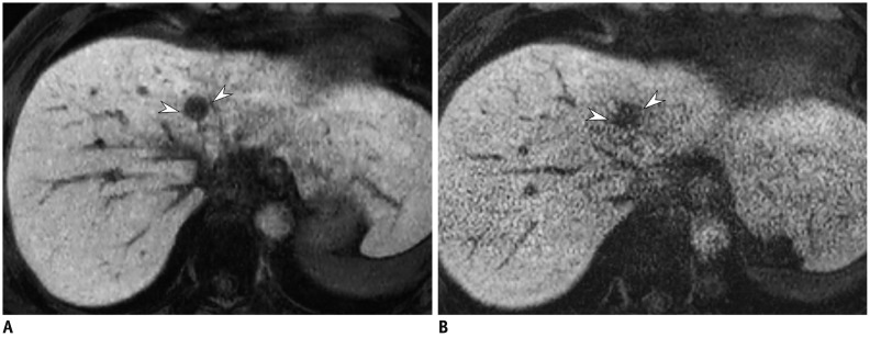
    1. Bashir MR, Castelli P, Davenport MS, Larson D, Marin D, Hussain HK, et al. Respiratory motion artifact affecting hepatic arterial phase MR imaging with gadoxetate disodium is more common in patients with a prior episode of arterial phase motion associated with gadoxetate disodium. Radiology. 2015;274:141–148. - PubMed
    1. Pietryga JA, Burke LM, Marin D, Jaffe TA, Bashir MR. Respiratory motion artifact affecting hepatic arterial phase imaging with gadoxetate disodium: examination recovery with a multiple arterial phase acquisition. Radiology. 2014;271:426–434. - PubMed
    1. Griswold MA, Jakob PM, Heidemann RM, Nittka M, Jellus V, Wang J, et al. Generalized autocalibrating partially parallel acquisitions (GRAPPA) Magn Reson Med. 2002;47:1202–1210. - PubMed
    1. Pruessmann KP, Weiger M, Scheidegger MB, Boesiger P. SENSE: sensitivity encoding for fast MRI. Magn Reson Med. 1999;42:952–962. - PubMed
    1. Hoogeveen RM, von Falkenhausen M, Gieseke J. Proceedings of the 12th scientific meeting and exhibition of the International Society for Magnetic Resonance in Medicine. Kyoto, Japan: 2004. May, Fast dynamic, high resolution contrast-enhanced MR angiography with CENTRA keyhole and SENSE; pp. 15–21.

Publication types