Multicenter Study
. 2019 Dec;20(12):1616-1626.
doi: 10.3348/kjr.2019.0363.

Intraindividual Comparison between Gadoxetate-Enhanced Magnetic Resonance Imaging and Dynamic Computed Tomography for Characterizing Focal Hepatic Lesions: A Multicenter, Multireader Study

Affiliations
Multicenter Study

Intraindividual Comparison between Gadoxetate-Enhanced Magnetic Resonance Imaging and Dynamic Computed Tomography for Characterizing Focal Hepatic Lesions: A Multicenter, Multireader Study

Chansik An et al. Korean J Radiol. 2019 Dec.

Abstract

Objective: To compare the diagnostic accuracy of dynamic computed tomography (CT) and gadoxetate-enhanced magnetic resonance imaging (MRI) for characterization of hepatic lesions by using the Liver Imaging Reporting and Data System (LI-RADS) in a multicenter, off-site evaluation.

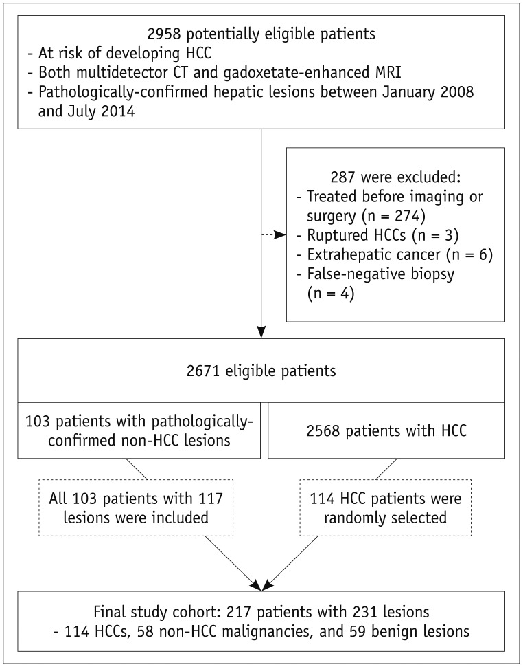
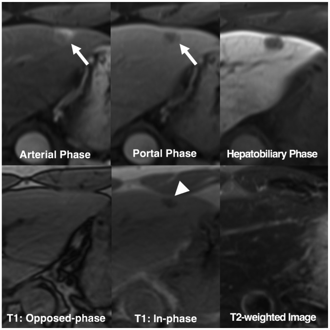
Materials and methods: In this retrospective multicenter study, we evaluated 231 hepatic lesions (114 hepatocellular carcinomas [HCCs], 58 non-HCC malignancies, and 59 benign lesions) confirmed histologically in 217 patients with chronic liver disease who underwent both gadoxetate-enhanced MRI and dynamic CT at one of five tertiary hospitals. Four radiologists at different institutes independently reviewed all MR images first and the CT images 4 weeks later. They evaluated the major and ancillary imaging features and categorized each hepatic lesion according to the LI-RADS v2014. Diagnostic performance was calculated and compared using generalized estimating equations.

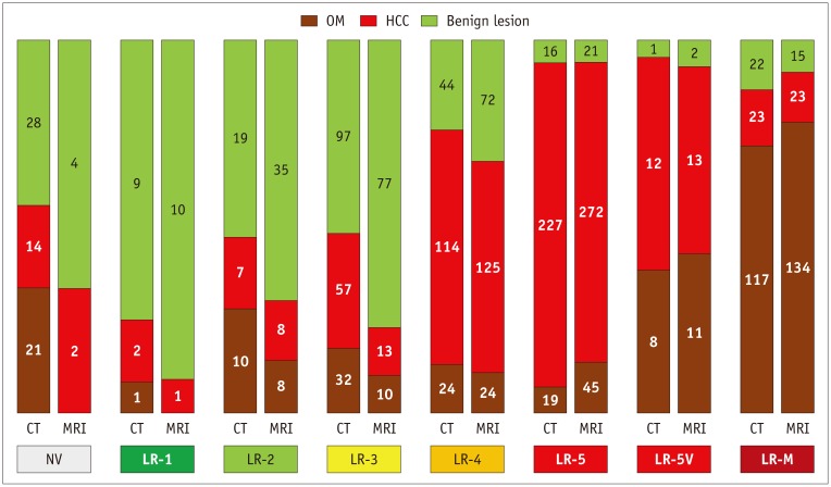
Results: MRI showed higher sensitivity and accuracy than CT for diagnosing hepatic malignancies; the pooled sensitivities, specificities, and accuracies for categorizing LR-5/5V/M were 59.0% vs. 72.4% (CT vs. MRI; p < 0.001), 83.5% vs. 83.9% (p = 0.906), and 65.3% vs. 75.3% (p < 0.001), respectively. CT and MRI showed comparable capabilities for differentiating between HCC and other malignancies, with pooled accuracies of 79.9% and 82.4% for categorizing LR-M, respectively (p = 0.139).

Conclusion: Gadoxetate-enhanced MRI showed superior accuracy for categorizing LR-5/5V/M in hepatic malignancies in comparison with dynamic CT. Both modalities had comparable accuracies for distinguishing other malignancies from HCC.

Keywords: Computed tomography; Contrast media; Data systems; Gadolinium ethoxybenzyl DTPA; Hepatocellular carcinoma; Magnetic resonance imaging.


Conflict of interest statement

The funder Bayer Korea Ltd. had no role in the study design; the collection, analysis, and interpretation of data; the writing of the report; or the decision to submit the manuscript for publication.

Figures

Fig. 1
Fig. 1. Flowchart illustrating subject selection.
CT = computed tomography, HCC = hepatocellular carcinoma, MRI = magnetic resonance imaging
Fig. 2
Fig. 2. Frequencies and proportions of HCC, OM, and benign lesions according to imaging modality and Liver Imaging Reporting and Data System category.
Numbers and areas of segments in each vertical bar indicate numbers and proportions of HCC, OM, and benign lesions, respectively. Pooled results from four reviewers are shown here (see Supplementary Table 1 for full results). NV = not visible, OM = other malignancies
Fig. 3
Fig. 3. Non-HCC malignancy with tumor in vein.
Diffuse hypervascular tumor with infiltrative margins is seen at right hemi-liver. Tumor also invades adjacent portal vein branch (P8), forming mass within vein (arrows). Two of our four reviewers categorized this mass as LR-5V, while other two assigned score of LR-M. Pathologic diagnosis obtained after biopsy was combined HCC-cholangiocarcinoma.
Fig. 4
Fig. 4. Pathologically-confirmed HCC categorized as LR-5 only with gadoxetate-enhanced MRI.
58-year-old female HBV carrier underwent both dynamic CT and gadoxetate-enhanced MRI with interval of 3 days. Her serum alpha-fetoprotein level was elevated at surveillance for HCC. On gadoxetate-enhanced MRI, all our reviewers found 1.8-cm nodule in right liver (arrow) with arterial hyperenhancement, portal washout appearance, and capsule appearance, and categorized this nodule as LR-5. However, on dynamic CT, only faint arterial hyperenhancement (arrowhead) was visualized at corresponding location. All reviewers categorized lesion as LR-3 or LR-4. HBV = hepatitis B virus
Fig. 5
Fig. 5. Benign lesion initially categorized as LR-5 but correctly downgraded by applying ancillary features.
52-year-old HBV carrier underwent gadoxetate-enhanced MRI after hepatic nodule was found on ultrasonography. 1.4-cm nodule in left liver shows arterial hyperenhancement (arrow) and washout appearance on portal phase (arrow). Two reviewers considered arterial enhancement as rim-like and categorized nodule as LR-M. Other reviewers initially categorized nodule as LR-5. Nodule shows signal drop from opposed-phase to in-phase of T1-weighted gradient-recalled echo sequence (arrowhead), indicating presence of intralesional iron deposits. Note that nodule shows isointensity on T2-weighted image. These features are uncommon finding in progressed HCCs. After applying these ancillary features, reviewers downgraded their categories to LR-4. This nodule was confirmed as angiomyolipoma after hepatic resection.

References

    1. Marrero JA, Kulik LM, Sirlin CB, Zhu AX, Finn RS, Abecassis MM, et al. Diagnosis, staging, and management of hepatocellular carcinoma: 2018 practice guidance by the American Association for the Study of Liver Diseases. Hepatology. 2018;68:723–750. - PubMed
    1. European Association for the Study of the Liver. EASL clinical practice guidelines: management of hepatocellular carcinoma. J Hepatol. 2018;69:182–236. - PubMed
    1. Omata M, Cheng AL, Kokudo N, Kudo M, Lee JM, Jia J, et al. Asia-Pacific clinical practice guidelines on the management of hepatocellular carcinoma: a 2017 update. Hepatol Int. 2017;11:317–370. - PMC - PubMed
    1. Korean Liver Cancer Study Group and Korean National Cancer Center. 2014 Korean Liver Cancer Study Group-National Cancer Center Korea practice guideline for the management of hepatocellular carcinoma. Korean J Radiol. 2015;16:465–522. - PMC - PubMed
    1. Kudo M, Trevisani F, Abou-Alfa GK, Rimassa L. Hepatocellular carcinoma: therapeutic guidelines and medical treatment. Liver Cancer. 2016;6:16–26. - PMC - PubMed

Publication types

MeSH terms

LinkOut - more resources