. 2019 Dec 19:8:e47839.
doi: 10.7554/eLife.47839.

De novo design of a homo-trimeric amantadine-binding protein

Affiliations

De novo design of a homo-trimeric amantadine-binding protein

Jooyoung Park et al. Elife. .

Abstract

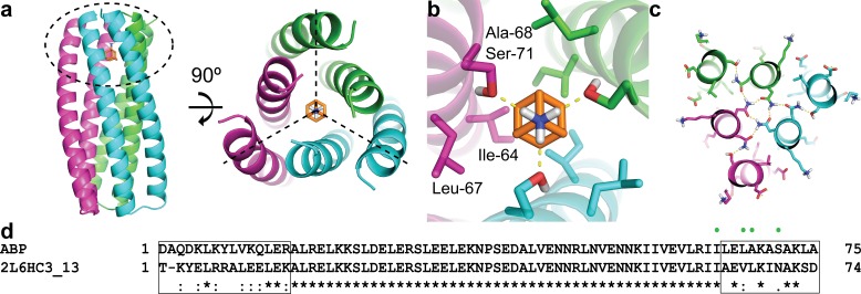
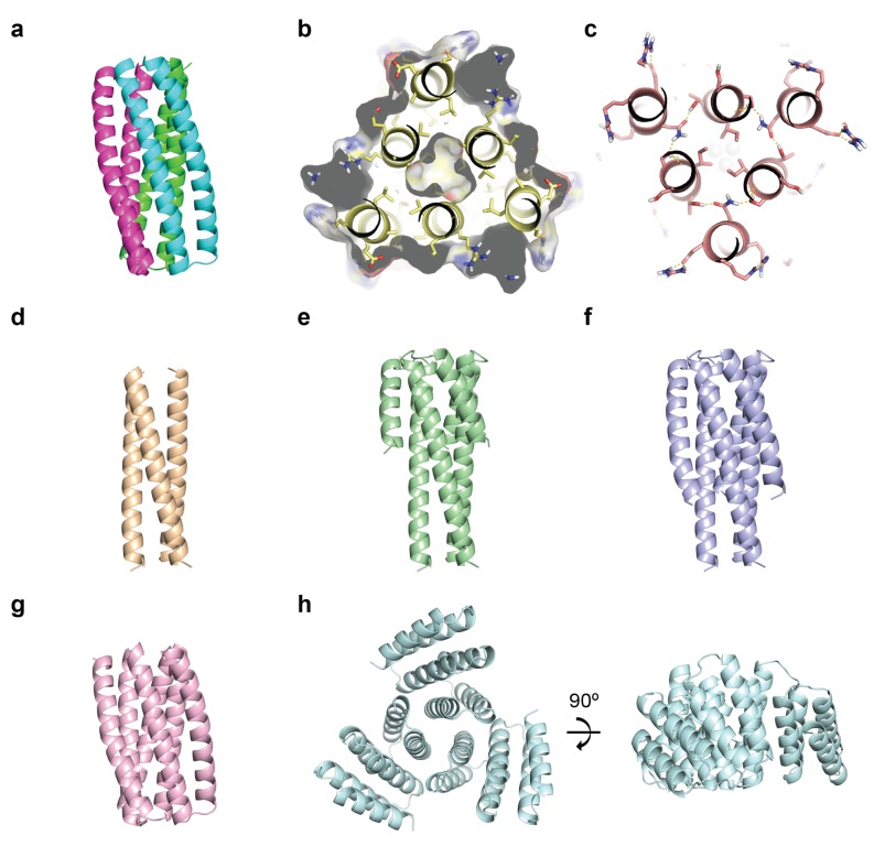
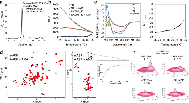
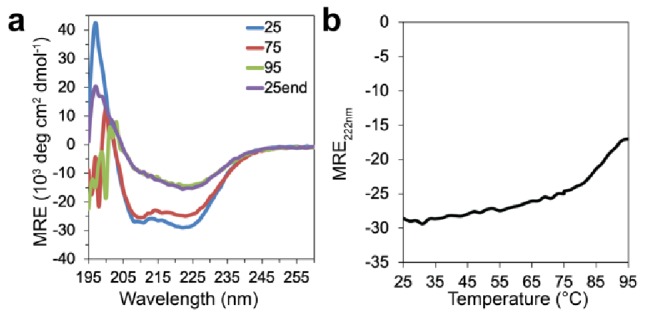
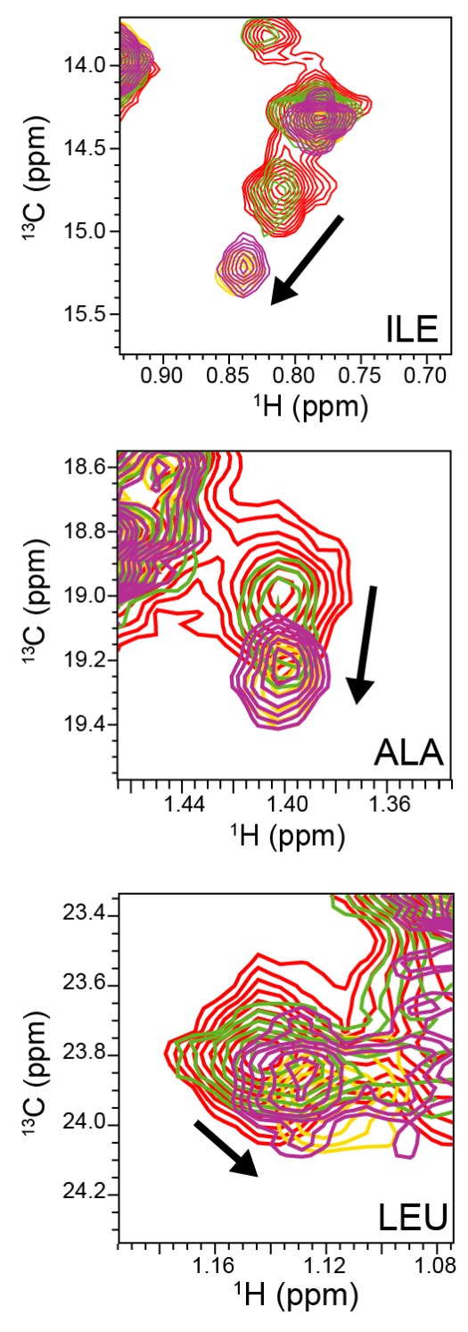
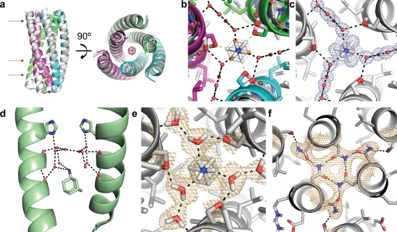
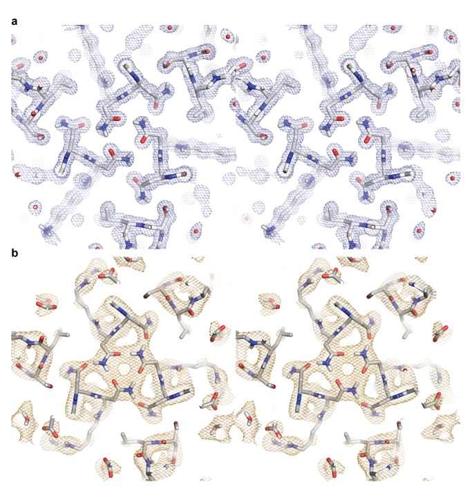
The computational design of a symmetric protein homo-oligomer that binds a symmetry-matched small molecule larger than a metal ion has not yet been achieved. We used de novo protein design to create a homo-trimeric protein that binds the C3 symmetric small molecule drug amantadine with each protein monomer making identical interactions with each face of the small molecule. Solution NMR data show that the protein has regular three-fold symmetry and undergoes localized structural changes upon ligand binding. A high-resolution X-ray structure reveals a close overall match to the design model with the exception of water molecules in the amantadine binding site not included in the Rosetta design calculations, and a neutron structure provides experimental validation of the computationally designed hydrogen-bond networks. Exploration of approaches to generate a small molecule inducible homo-trimerization system based on the design highlight challenges that must be overcome to computationally design such systems.

Keywords: E. coli; Rosetta; amantadine; de novo protein design; molecular biophysics; structural biology; symmetry.


Conflict of interest statement

JP, SB, KW, GO, DB JP, SEB, KYW, GO, and DB have filed a provisional application based for "Amantadine Binding Protein" (Application # 62/834,592). BS, AM, WD, NS, MC, DM No competing interests declared

Figures

Figure 1.
Figure 1.. Computational design methodology.
(a) The homo-trimeric scaffold was designed to bind amantadine such that the C3 axes of the protein and the small molecule are aligned. Amantadine is colored orange and each monomer of ABP is colored magenta, green, or cyan. The amantadine binding site is highlighted by a dashed oval. (b) The binding pocket in ABP was designed to have polar serine residues (Ser-71) that hydrogen-bond (yellow dashed lines) to the amino group of amantadine and nonpolar residues (Ile-64, Leu-67, and Ala-68) to complement the shape of the hydrophobic moiety of amantadine. (c) The design model contains hydrogen-bond networks that specify the trimeric assembly of ABP. (d) A sequence alignment of ABP and 2L6HC3_13 is shown, with mutated regions shown in black boxes. Both sequences are preceded by a five-residue GHSMG pre-sequence (not shown) that result from the cloning strategy. The residues highlighted in (b) are annotated by green circles.
Figure 1—figure supplement 1.
Figure 1—figure supplement 1.. ABP variant designs.
(a) The original ABP design is shown colored in magenta, green, or cyan. (b) Ala/Ser mutations were introduced in the core of the helical bundle in order to reduce the trimeric interface. (c) HBNet was used to incorporate more polar residues at the monomer-monomer interfaces that could make hydrogen-bond interactions (yellow dashed lines). (d-f) The outer helices were truncated to various lengths and the identities of surface-exposed positions redesigned to be polar. (g) The helical bundle was shortened by removing a heptad repeat. (h) The helical bundle was shortened to just the amantadine-binding site, and helical repeats were fused onto the core bundle in order to stabilize the structure of the reduced core.
Figure 2.
Figure 2.. Binding characterization of amantadine to ABP.
(a) SEC chromatogram monitoring absorbance at 280 nm (mAU) and estimated molecular mass (from MALS). (b) Apo-ABP (orange, open circle) exhibits a high initial fluorescence signal that is lowered in the presence of amantadine (orange, solid circle). As expected, 2LC3H6_13 (blue, open triangle) and 2LC3H6_13 plus amantadine (blue, solid triangle) exhibit a very low initial fluorescence signal and overlap almost identically. (c) The CD spectrum of ABP at 25°C, 75°C, 95°C, and 25°C after heating and cooling. The CD spectrum of ABP at 25°C suggests an all ɑ-helical structure that remains fairly stable up to 75°C. (d) 2D amide 1H-15N HMQC spectra (left) and 2D methyl 1H-13C HMQC spectra (right) of 250 μM apo-ABP (red) or ABP in the presence of 2 mM amantadine (blue) recorded at 800 MHz, 37°C. Titration of amantadine leads to significant changes in the ABP NMR spectra (arrows). To the right of the 2D methyl methyl 1H-13C HMQC spectra an inset of dissociation constant estimate through conventional fraction bound analysis is shown for the affected ILE methyl group, with an estimated KD of <55 μM. (e) NMR line shape fitting of ABP throughout the NMR titration with amantadine performed in the program TITAN using a two-state binding model for the affected ILE methyl group. The NMR data (gray) are shown versus the TITAN fit (magenta).
Figure 2—figure supplement 1.
Figure 2—figure supplement 1.. CD spectrum of ABP in the presence amantadine.
(a) The CD spectrum of ABP in the presence of 5 mM amantadine at 25°C, 75°C, 95°C, and 25°C after heating and cooling suggests that the thermal stability of ABP is not significantly affected by the presence of amantadine. (b) CD temperature melt monitoring absorption at 222 nm for ABP in the presence of 5 mM amantadine.
Figure 2—figure supplement 2.
Figure 2—figure supplement 2.. NMR titration of ABP with amantadine.
Representative view of 2D 1H-13C methyl SOFAST-HMQC spectra of select ABP methyl groups without amantadine (red) and with amantadine at ABP:amantadine molar ratios of 1:0.58 (green), 1:1.35 (purple), and 1:2.47 (yellow). Arrows indicate significant chemical shift deviations in ABP methyl groups upon complex formation with amantadine. NMR experiments were recorded with 118 μM ABP at 800 MHz, 37°C.
Figure 3.
Figure 3.. Structural characterization of the ABP-amantadine interaction.
(a) The high-resolution X-ray structure (white) and neutron structure (gray) of ABP in complex with amantadine are very close to the computational model (magenta, green, and cyan) (RMSD of 0.63 Å and 0.59 Å, respectively). The blue arrow indicates the amantadine binding site shown in (c,e), and the red arrows indicate the hydrogen bond networks, one of which is shown in (f). (b) A zoomed in overlay of the X-ray structure (white) and the design model (colored) reveal a shift in the helices within the amantadine-binding region, accompanied by a ~ 60° rotation of amantadine and the presence of water molecules that that mediate hydrogen bonding to Ser-71. Yellow dashed lines show direct hydrogen bonds to Ser-71 in the design model and black dashed lines show the hydrogen bonds observed in the X-ray structure. (c) Clear electron density can be observed for amantadine and ordered water molecules in the binding site of ABP (2Fo - Fc map contoured at 1.0σ). Water-mediated hydrogen bonds are observed between Ser-71 and the amino group of amantadine (black dashed lines). (d) The crystal structure (pale green) of amantadine bound to the influenza M2 protein through water-mediated hydrogen bonds (image generated from PDB: 6BKK [Thomaston et al., 2018]). (e) The nuclear scattering length density map shows the positions of deuterium atoms, including two ordered water molecules that mediate the hydrogen-bond network between Ser-71 and amantadine (2Fo - Fc contoured at 1.0σ). Hydrogen bonds are shown as black dashed lines. (f) Clear nuclear scattering length density can be observed for residues involved in the designed hydrogen-bond networks (black dashed lines) in ABP (2Fo - Fc map contoured at 1.0σ).
Figure 3—figure supplement 1.
Figure 3—figure supplement 1.. Stereo images of the electron and neutron length scattering density maps for a hydrogen bond network in ABP.
(a) The 2Fo - Fc electron density map contoured at 1.0σ is colored in light blue. (b) The 2Fo - Fc neutron density length scattering map contoured at 1.0σ is colored in light orange.

References

    1. Adams PD, Afonine PV, Bunkóczi G, Chen VB, Davis IW, Echols N, Headd JJ, Hung LW, Kapral GJ, Grosse-Kunstleve RW, McCoy AJ, Moriarty NW, Oeffner R, Read RJ, Richardson DC, Richardson JS, Terwilliger TC, Zwart PH. PHENIX: a comprehensive Python-based system for macromolecular structure solution. Acta Crystallographica Section D Biological Crystallography. 2010;66:213–221. doi: 10.1107/S0907444909052925. - DOI - PMC - PubMed
    1. Afonine PV, Mustyakimov M, Grosse-Kunstleve RW, Moriarty NW, Langan P, Adams PD. Joint X-ray and neutron refinement with phenix.refine. Acta Crystallographica. Section D, Biological Crystallography. 2010;66:1153–1163. doi: 10.1107/S0907444910026582. - DOI - PMC - PubMed
    1. Arzt S, Campbell JW, Harding MM, Hao Q, Helliwell JR. LSCALE-the new normalization, scaling and absorption correction program in the Daresbury laue software suite. Journal of Applied Crystallography. 1999;32:554–562. doi: 10.1107/S0021889898015350. - DOI
    1. Bale JB, Gonen S, Liu Y, Sheffler W, Ellis D, Thomas C, Cascio D, Yeates TO, Gonen T, King NP, Baker D. Accurate design of megadalton-scale two-component icosahedral protein complexes. Science. 2016;353:389–394. doi: 10.1126/science.aaf8818. - DOI - PMC - PubMed
    1. Boyken SE, Chen Z, Groves B, Langan RA, Oberdorfer G, Ford A, Gilmore JM, Xu C, DiMaio F, Pereira JH, Sankaran B, Seelig G, Zwart PH, Baker D. De novo design of protein homo-oligomers with modular hydrogen-bond network-mediated specificity. Science. 2016;352:680–687. doi: 10.1126/science.aad8865. - DOI - PMC - PubMed

Publication types