. 2020 Mar 7;8(5):1256-1269.
doi: 10.1039/c9bm01894h. Epub 2019 Dec 19.

Hierarchically structured hydrogels utilizing multifunctional assembling peptides for 3D cell culture

Affiliations

Hierarchically structured hydrogels utilizing multifunctional assembling peptides for 3D cell culture

Amber M Hilderbrand et al. Biomater Sci. .

Abstract

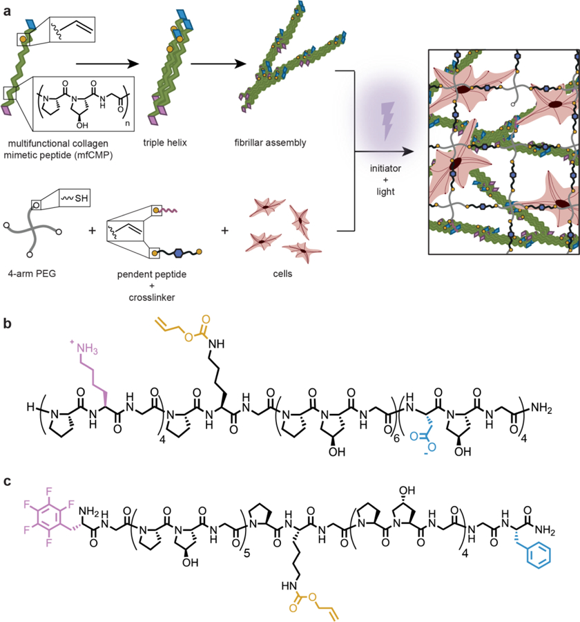
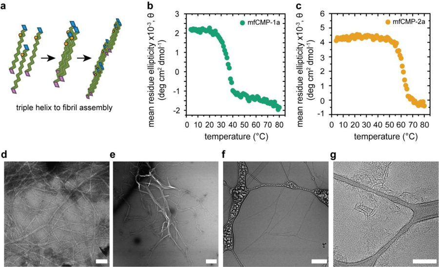
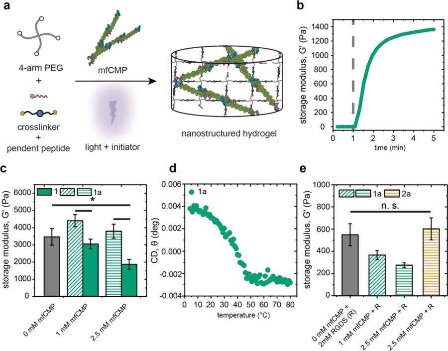
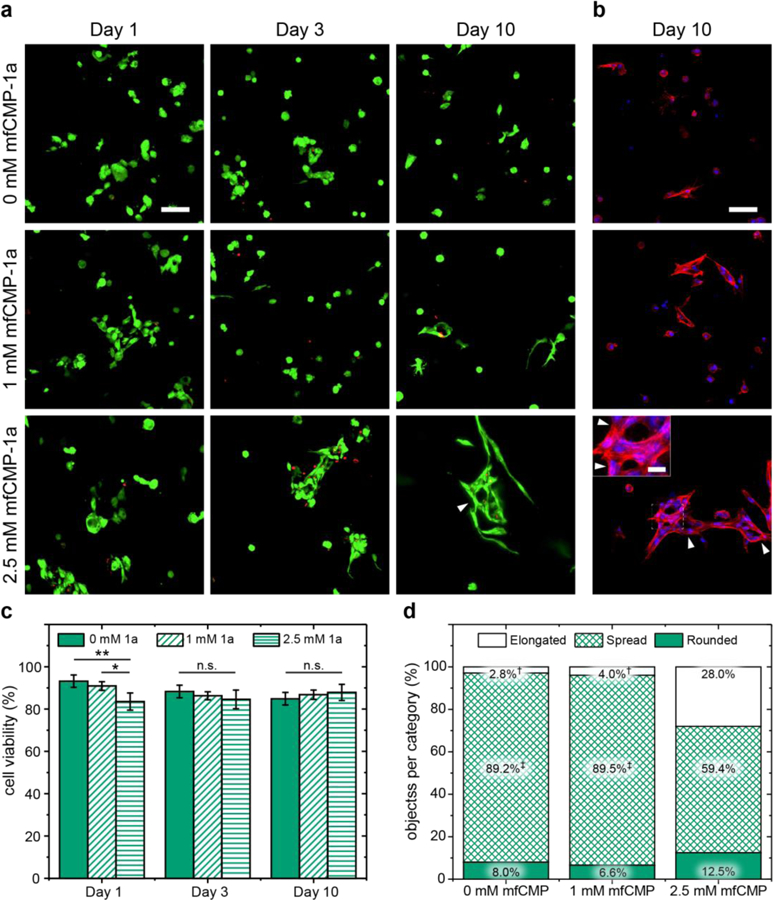
Approaches for the creation of soft materials, particularly hydrogels, with hierarchical structure are of interest in a variety of applications owing to their unique properties. In the context of tissue mimics, hydrogels with multiscale structures more accurately capture the complexities of tissues within the body (e.g., fibrous collagen-rich microenvironments). However, cytocompatible fabrication of such materials with hierarchical structures and independent control of mechanical and biochemical properties remains challenging and is needed for probing and directing cell-microenvironment interactions for three-dimensional (3D) cell encapsulation and culture applications. To address this, we have designed innovative multifunctional assembling peptides: these unique peptides contain a core block that mimics the structure of collagen for achieving relevant melting temperatures; 'sticky' ends to promote assembly of long fibrils; and a biocompatible reactive handle that is orthogonal to assembly to allow the formation of desired multiscale structures and their subsequent rapid, light-triggered integration within covalently crosslinked synthetic hydrogels. Nano- to micro-fibrils were observed to form in physiologically-relevant aqueous solutions, where both underlying peptide chemical structure and assembly conditions were observed to impact the resulting fibril sizes. These assembled structures were 'locked' into place and integrated as linkers within cell-degradable, bioactive hydrogels formed with photoinitiated thiol-ene 'click' chemistry. Hydrogel compositions were identified for achieving robust mechanical properties like those of soft tissues while also retaining higher ordered structures after photopolymerization. The utility of these innovative materials for 3D cell culture was demonstrated with human mesenchymal stem cells, where cell morphologies reminiscent of responses to assembled native collagen were observed now with a fully synthetic material. Using a bottom-up approach, a new materials platform has been established that combines the advantageous properties of covalent and assembling chemistries for the creation of synthetic hydrogels with controllable nanostructure, mechanical properties, and biochemical content.


Figures

Fig. 1:
Fig. 1:
Approach to creating robust hierarchically-structured hydrogels. a) Multifunctional CMPs (mfCMPs) were designed for assembly and subsequent integration within covalently crosslinked hydrogels formed by light-triggered thiol–ene click chemistry, with a 4-arm PEG-SH macromer, cell-degradable peptide linker, photoinitiator, and integrin-binding pendant peptide. This approach allows for the encapsulation and 3D culture of cells within a robust, synthetic hydrogel with multiscale structures. Two mfCMP designs were established: b) mfCMP-1a (‘sticky’ ends lysine (pink) and aspartic acid (blue); reactive handle alloc-protected lysine (yellow)) and c) mfCMP-2a (aromatic stacking ends pentafluorophenylalanine (pink) and phenylalanine (blue)).
Fig. 2:
Fig. 2:
Assembly of mfCMP into triple helices and fibrils in solution. a) Schematic: three peptide strands assemble into triple helices as a result of hydrogen bonding, and these subsequently assemble into fibrils, facilitated by end-to-end interactions imparted by the charged or aromatic amino acid substitutions. CD of b) mfCMP-1a and c) mfCMP-2a measuring the ellipticity of the sample at a wavelength of 225 nm over a range of temperatures to determine the melting temperature of each peptide. TEM of d) mfCMP-1a and e) mfCMP-2a after assembly at 1 mM concentration and negatively stained with 2% uranyl acetate. Scale bars = 500 nm. Cryo-TEM of f) mfCMP-1a and g) mfCMP-2a assembled at higher concentrations and diluted to 1 mM immediately before preparing samples, mirroring conditions used for hydrogel formation. Scale bars = 200 nm.
Fig. 3:
Fig. 3:
Mechanical properties of nanostructured hydrogels containing mfCMPs. a) Schematic of approach for incorporation of assembled mfCMPs within hydrogels using K(alloc) handle that is orthogonal to assembly: mfCMPs were assembled in solution for 48 h, diluted into a hydrogel precursor solution, and immediately irradiated in situ to form robust, nanostructured hydrogels. b) In situ oscillatory rheometry was used to monitor the rapid formation of these hydrogels (dashed gray line indicates when irradiation commenced; here, hydrogel formed with 2.5 mM mfCMP-1a as representative example), and their resulting mechanical properties compared. Hydrogels formed with c) increasing amounts of mfCMPs with or without an alloc group (mfCMP-1a or mfCMP-1, respectively). d) CD on an in situ hydrogel containing 2.5 mM mfCMP-1a to determine the melting temperature of assembled mfCMPs within the covalently crosslinked network. e) Moduli of hydrogels formed with mfCMPs and integrin binding peptide RGDS equilibrated in DPBS at 37 °C, which were the formulations used in 3D cell culture studies. The data shown illustrate the mean with error bars showing the standard error (n ≥ 3; * p < 0.05).
Fig. 4:
Fig. 4:
Effects of nanostructured mfCMP-1a hydrogels on hMSC viability and morphology in 3D cell culture. Representative maximum intensity projections from confocal z-stacks (200 μm thick) of hMSCs encapsulated within hydrogels without mfCMP-1a (0 mM), at a low concentration of mfCMP-1a (1 mM), or at a high concentration (2.5 mM) of mfCMP-1a for a) Live/Dead cytotoxicity assay (live cells, green; dead cell nuclei, red) and b) fixed stained samples at day 10 (F-actin cytoskeleton, red; cell nuclei, blue). Scale bars: large images = 100 μm, inset image = 25 μm. Arrowheads note unique ‘holes’ that were consistently observed within cell clusters when cultured in 2.5 mM mfCMP-1a hydrogels. From these images, c) cell viability was quantified (error bars represent the standard error around the mean; n = 3 hydrogels, > 250 cells counted per condition; *p < 0.05, **p < 0.01). d) Cell morphology was analyzed through quantification of 3D shape factor (Ψ) of individual or clusters of cells at day 10 with a focus on analysis of cell elongation († indicates a significant difference in the percent of elongated objects relative to the experimental condition 2.5 mM mfCMP-1a, p < 0.05; ‡ indicates a significant difference in the percentage of spread objects relative to the experimental condition 2.5 mM mfCMP-1a, p < 0.05); full set of quantitative results and statistics are available in Tables S1–S4).

References

    1. Hilderbrand AM, Ovadia EM, Rehmann MS, Kharkar PM, Guo C and Kloxin AM, Current Opinion in Solid State and Materials Science, 2016, 20, 212–224. - PMC - PubMed
    1. Brown TE and Anseth KS, Chemical Society Reviews, 2017, 46, 6532–6552. - PMC - PubMed
    1. Murphy WL, McDevitt TC and Engler AJ, Nature Materials, 2014, 13, 547–557. - PMC - PubMed
    1. Rose JC and De Laporte L, Advanced Healthcare Materials, 2018, 7, 1701067. - PubMed
    1. Abraham LC, Zuena E, Perez-Ramirez B and Kaplan DL, Journal of Biomedical Materials Research Part B: Applied Biomaterials, 2008, 87B, 264–285. - PubMed