

Mapping Enzyme Activity on Tissue by Functional Mass Spectrometry Imaging
- PMID: 31854493
- PMCID: PMC7106485
- DOI: 10.1002/anie.201911390
Mapping Enzyme Activity on Tissue by Functional Mass Spectrometry Imaging
Abstract
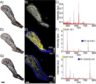
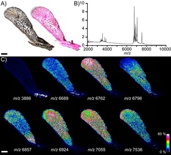
Enzymes are central components of most physiological processes, and are consequently implicated in various pathologies. High-resolution maps of enzyme activity within tissues therefore represent powerful tools for elucidating enzymatic functions in health and disease. Here, we present a novel mass spectrometry imaging (MSI) method for assaying the spatial distribution of enzymatic activity directly from tissue. MSI analysis of tissue sections exposed to phospholipid substrates produced high-resolution maps of phospholipase activity and specificity, which could subsequently be compared to histological images of the same section. Functional MSI thus represents a new and generalisable method for imaging biological activity in situ.
Keywords: PLA2; enzymes; functional assays; lipids; mass spectrometry imaging.
© 2019 The Authors. Published by Wiley-VCH Verlag GmbH & Co. KGaA.
Conflict of interest statement
The authors declare no conflict of interest.
Figures

Similar articles
-
Characterization and immunological comparison of isoenzymes of phospholipases A2 from snake venoms of different genera and families.Biochem Int. 1991 Dec;25(6):1003-11. Biochem Int. 1991. PMID: 1810246
-
The biflavonoid morelloflavone inhibits the enzymatic and biological activities of a snake venom phospholipase A2.Chem Biol Interact. 2014 Sep 5;220:94-101. doi: 10.1016/j.cbi.2014.06.015. Epub 2014 Jul 1. Chem Biol Interact. 2014. PMID: 24995575
-
Functional mass spectrometry imaging maps phospholipase-A2 enzyme activity during osteoarthritis progression.Theranostics. 2023 Aug 21;13(13):4636-4649. doi: 10.7150/thno.86623. eCollection 2023. Theranostics. 2023. PMID: 37649605 Free PMC article.
-
Calcium-independent membrane damage by venom phospholipases A2.Protein Pept Lett. 2009;16(8):877-86. doi: 10.2174/092986609788923392. Protein Pept Lett. 2009. PMID: 19689413 Review.
-
Cytotoxicity of snake venom enzymatic toxins: phospholipase A2 and l-amino acid oxidase.Biochem Soc Trans. 2020 Apr 29;48(2):719-731. doi: 10.1042/BST20200110. Biochem Soc Trans. 2020. PMID: 32267491 Free PMC article. Review.
Cited by
-
Mapping enzyme activity in living systems by real-time mid-infrared photothermal imaging of nitrile chameleons.Nat Methods. 2024 Feb;21(2):342-352. doi: 10.1038/s41592-023-02137-x. Epub 2024 Jan 8. Nat Methods. 2024. PMID: 38191931 Free PMC article.
-
Multiplex enzyme activity imaging by MALDI-IMS of substrate library conversions.Sci Rep. 2020 Sep 23;10(1):15522. doi: 10.1038/s41598-020-72436-2. Sci Rep. 2020. PMID: 32968143 Free PMC article.
-
High-resolution imaging and identification of biomolecules using Nano-DESI coupled to ion mobility spectrometry.Anal Chim Acta. 2021 Nov 22;1186:339085. doi: 10.1016/j.aca.2021.339085. Epub 2021 Sep 21. Anal Chim Acta. 2021. PMID: 34756271 Free PMC article.
-
Insights into how development and life-history dynamics shape the evolution of venom.Evodevo. 2021 Jan 7;12(1):1. doi: 10.1186/s13227-020-00171-w. Evodevo. 2021. PMID: 33413660 Free PMC article. Review.
-
Ozone-enabled fatty acid discovery reveals unexpected diversity in the human lipidome.Nat Commun. 2023 Jul 4;14(1):3940. doi: 10.1038/s41467-023-39617-9. Nat Commun. 2023. PMID: 37402773 Free PMC article.
References
-
- Mohamed M. M., Sloane B. F., Nat. Rev. Cancer 2006, 6, 764–775. - PubMed
Publication types
MeSH terms
Substances
Grants and funding
LinkOut - more resources
Full Text Sources
Other Literature Sources
