Review
. 2020 Feb 19;31(2):375-395.
doi: 10.1021/acs.bioconjchem.9b00758. Epub 2020 Jan 6.

Image-Guided Surgery: Are We Getting the Most Out of Small-Molecule Prostate-Specific-Membrane-Antigen-Targeted Tracers?

Affiliations
Review

Image-Guided Surgery: Are We Getting the Most Out of Small-Molecule Prostate-Specific-Membrane-Antigen-Targeted Tracers?

Albertus Wijnand Hensbergen et al. Bioconjug Chem. .

Abstract

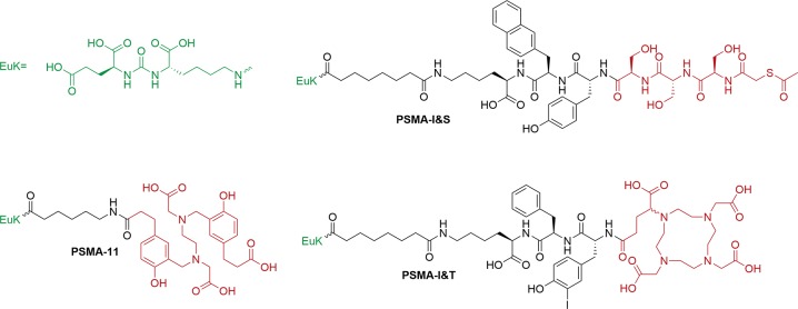
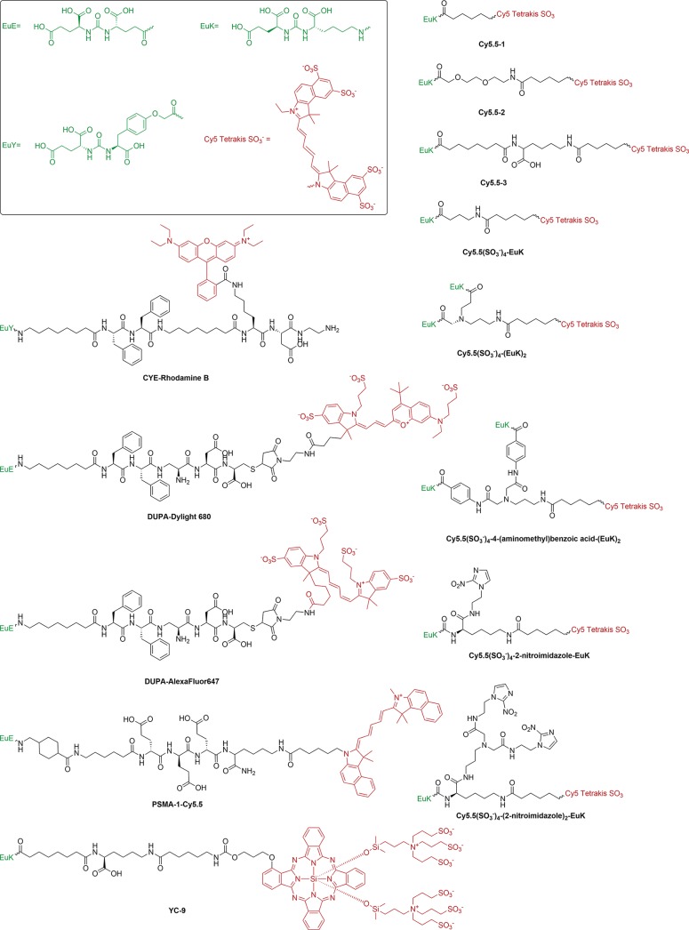
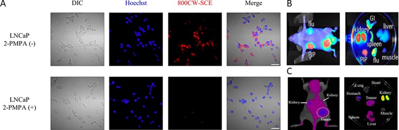
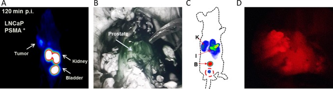
Expressed on virtually all prostate cancers and their metastases, the transmembrane protein prostate-specific membrane antigen (PSMA) provides a valuable target for the imaging of prostate cancer. Not only does PSMA provide a target for noninvasive diagnostic imaging, e.g., PSMA-positron emission tomography (PSMA-PET), it can also be used to guide surgical resections of PSMA-positive lesions. The latter characteristic has led to the development of a plethora of PSMA-targeted tracers, i.e., radiolabeled, fluorescent, or hybrid. With image-guided surgery applications in mind, this review discusses these compounds based on clinical need. Here, the focus is on the chemical aspects (e.g., imaging label, spacer moiety, and targeting vector) and their impact on in vitro and in vivo tracer characteristics (e.g., affinity, tumor uptake, and clearance pattern).


Conflict of interest statement

The authors declare the following competing financial interest(s): F. W. B. van Leeuwen is Chief Innovation Officer at ORSI Academy, and H.-J. Wester is founder and shareholder of Scintomics GmbH.

Figures

Figure 1
Figure 1
(A) Schematic cross-section view of PSMA illustrating intrinsic interactions between the three critical tracer components (targeting vector, spacer, and imaging flag) and the protein’s primary and secondary binding site. (B) Depiction of anatomical relevance in PCa surgery: bladder (in blue) relative to liver (red), kidneys (blue), lymph nodes (green), and prostate (pink). (C) Typical intraoperative sight during radical prostatectomy using nerve-sparing surgery.
Figure 2
Figure 2
Typical clinical workflow of radioguided surgery: (A) preoperative 68Ga-PSMA-11 PET/CT followed by (B) preoperative 99mTc-PSMA I&S SPECT/CT; (C) intraoperative guidance combined with minimally invasive robot-assisted laparoscopic surgery using a DROP-IN γ-probe with 99mTc-PSMA I&S. (Reprinted from van Leeuwen et al. with permission from Wolters Kluwer Health, Inc.).
Scheme 1
Scheme 1. Chemical Structures of PSMA-Targeted Tracers for PET and SPECT Imaging
Targeting moiety (green) and imaging flag (red) have been colored accordingly.
Scheme 2
Scheme 2. Chemical Structures of Visible to Far-Red Fluorescent Imaging Agents for PSMA-Targeted Fluorescence-Guided Surgery
Targeting vector (green) and imaging label (red) were colored accordingly.
Figure 3
Figure 3
(A) In vitro uptake of IRDye800CW-SCE in PSMA-positive LNCaP cells (top) with blocking by 2-PMPA (bottom) (adapted from Matusoka et al. with permission from Elsevier). Example of in vivo imaging and typical biodistribution for (B) IRDye800CW-1 (adapted from Chen et al.) and (C) Cy5.5(SO3)4-EuK (adapted from Kwon et al.)
Scheme 3
Scheme 3. Chemical Structures of Near-Infrared Imaging Agents for PSMA-Targeted Fluorescence-Guided Surgery
Targeting vector (green) and imaging label (red) were colored accordingly.
Scheme 4
Scheme 4. Chemical Structures of Hybrid Tracers for PSMA-Targeted Image-Guided Surgery
Targeting vector (green) and imaging label (red) were colored accordingly.
Figure 4
Figure 4
(A) Maximum-intensity projections of small-animal PET imaging of athymic nude mice bearing LNCaP tumors using 0.5 nmol 68Ga-HBED-CC-IRDye800CW. (B) Use of HBED-CC-IRDye800CW in vivo for fluorescence imaging with a Da Vinci Firefly laparoscope (1 mg, 30 μg/kg; both adapted from Baranski et al.; in JNM). (C) Maximum-intensity projections of small-animal SPECT imaging of BALB/c nude mice bearing LNCaP tumors using 1.0 nmol 99mTc-EuK-(SO3)Cy5-COOH (adapted from Hensbergen et al. in JNM). (D) Use of 99mTc-EuK-(SO3)Cy5-COOH (100 μg, 2.85 μg/kg) for in vivo fluorescence imaging with a prototype Karl Storz camera that allows Cy5 imaging.

References

    1. Bray F.; Ferlay J.; Soerjomataram I.; Siegel R. L.; Torre L. A.; Jemal A. (2018) Global cancer statistics 2018: GLOBOCAN estimates of incidence and mortality worldwide for 36 cancers in 185 countries. Ca-Cancer J. Clin. 68, 394–424. 10.3322/caac.21492. - DOI - PubMed
    1. Silver D. A.; Pellicer I.; Fair W. R.; Heston W. D.; Cordon-Cardo C. (1997) Prostate-specific membrane antigen expression in normal and malignant human tissues. Clin. Cancer Res. 3, 81–5. - PubMed
    1. Jin J. K.; Dayyani F.; Gallick G. E. (2011) Steps in prostate cancer progression that lead to bone metastasis. Int. J. Cancer 128, 2545–61. 10.1002/ijc.26024. - DOI - PMC - PubMed
    1. Maurer T.; Eiber M.; Schwaiger M.; Gschwend J. E. (2016) Current use of PSMA-PET in prostate cancer management. Nat. Rev. Urol. 13, 226–35. 10.1038/nrurol.2016.26. - DOI - PubMed
    1. Schwarzenböck S. M.; Rauscher I.; Blümel C.; Fendler W. P.; Rowe S. P.; Pomper M. G.; Afshar-Oromieh A.; Herrmann K.; Eiber M. (2017) PSMA Ligands for PET Imaging of Prostate Cancer. J. Nucl. Med. 58, 1545–1552. 10.2967/jnumed.117.191031. - DOI - PubMed

Publication types

MeSH terms