

Potassium iodide enhances the photobactericidal effect of methylene blue on Enterococcus faecalis as planktonic cells and as biofilm infection in teeth
- PMID: 31855718
- PMCID: PMC6947667
- DOI: 10.1016/j.jphotobiol.2019.111730
Potassium iodide enhances the photobactericidal effect of methylene blue on Enterococcus faecalis as planktonic cells and as biofilm infection in teeth
Erratum in
-
Corrigendum to "Potassium iodide enhances the photobactericidal effect of methylene blue on Enterococcus faecalis as planktonic cells and as biofilm infection in teeth" [J Photochem Photobiol B 203(2020) 1-11/111730].J Photochem Photobiol B. 2020 Jun;207:111892. doi: 10.1016/j.jphotobiol.2020.111892. Epub 2020 May 11. J Photochem Photobiol B. 2020. PMID: 32408121 No abstract available.
Abstract
Objective: To explore the effectiveness, biosafety, photobleaching and mechanism of antimicrobial photodynamic therapy (aPDT) using methylene blue (MB) plus potassium iodide (KI), for root canal infections.
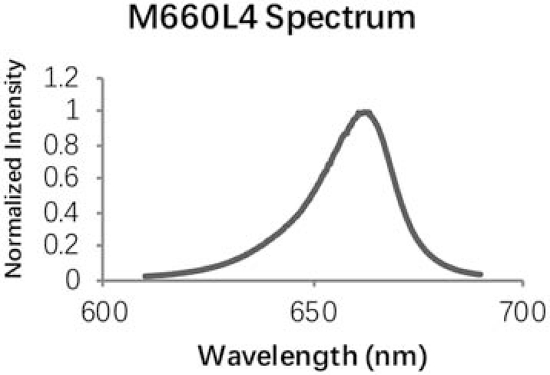
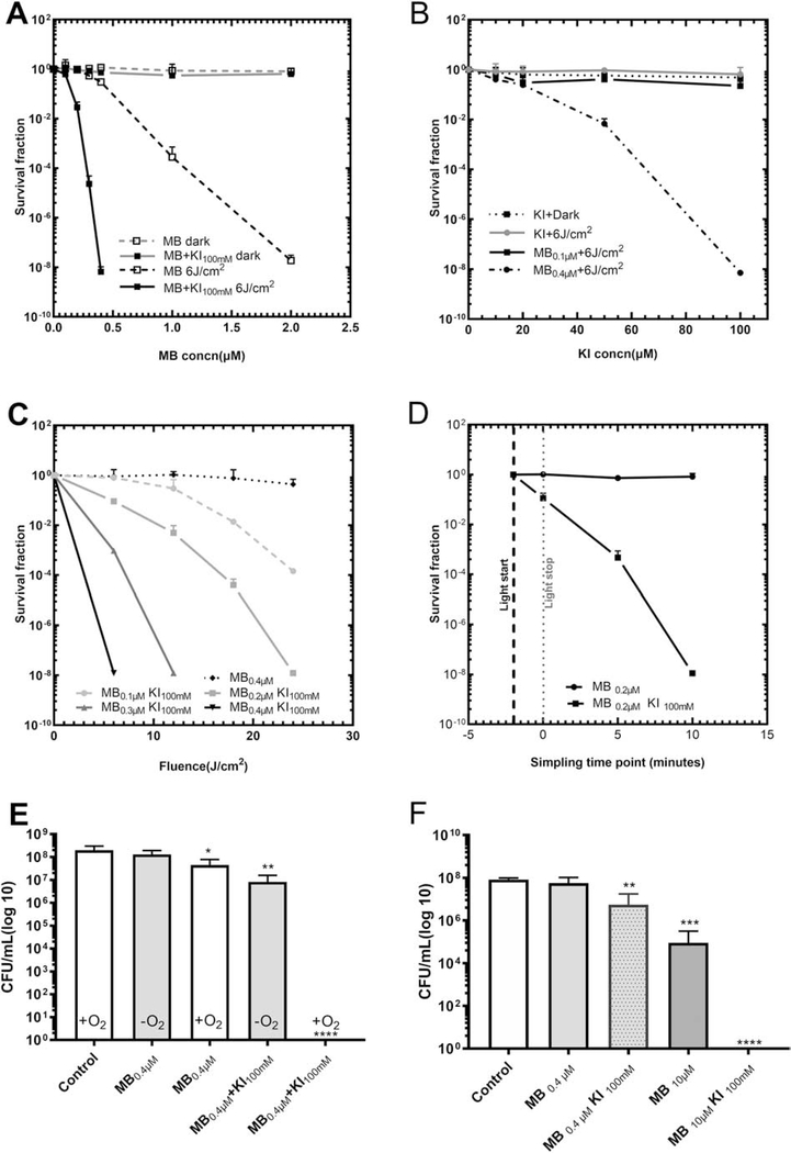
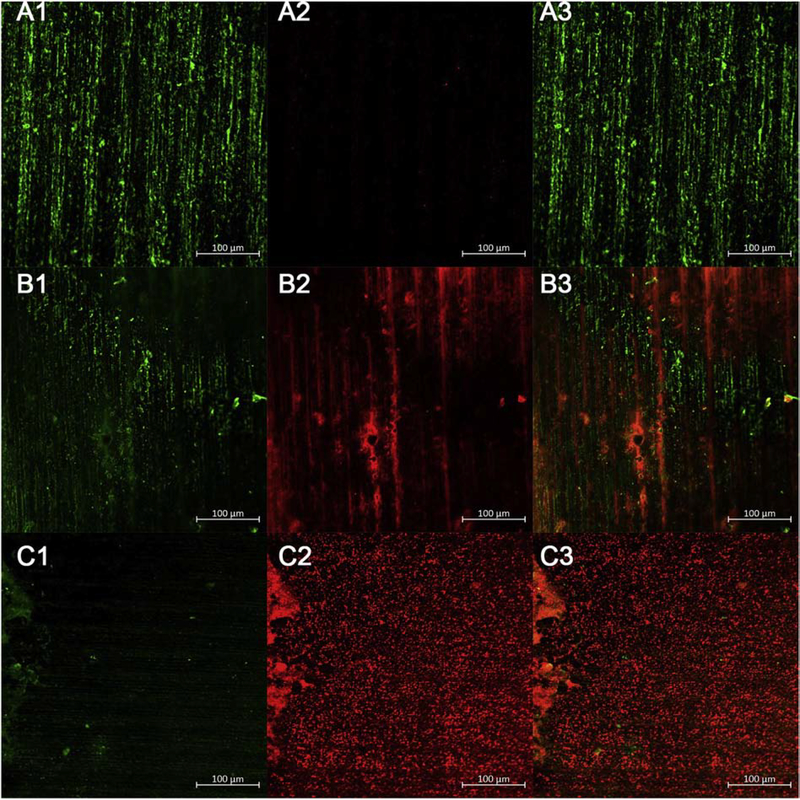
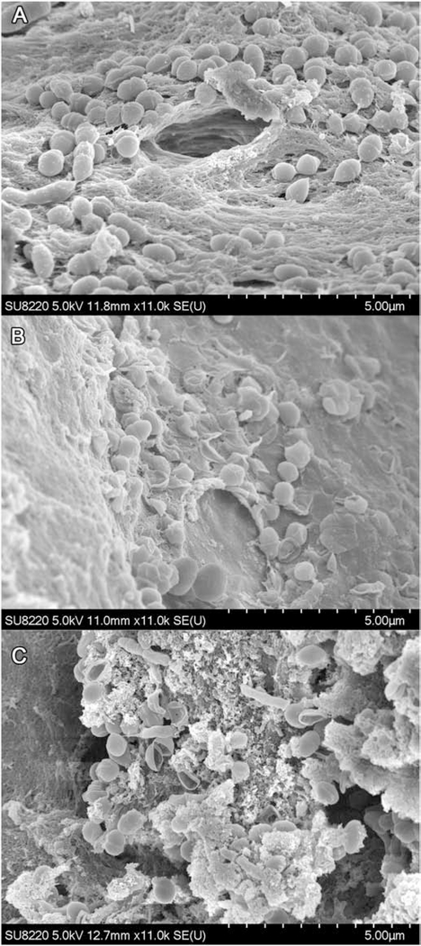
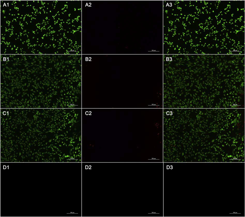
Methods: Different combinations and concentrations of MB, KI and 660 nm LED light were used against E. faecalis in planktonic and in biofilm states by colony-forming unit (CFU), confocal laser scanning microscopy (CLSM), scanning electron microscopy (SEM). Human gingival fibroblasts (HGF) were used for safety testing by Cell Counting Kit-8 (CCK8) and fluorescence microscopy (FLM). The photobleaching effect and mechanisms were analyzed.
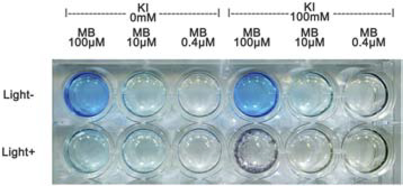
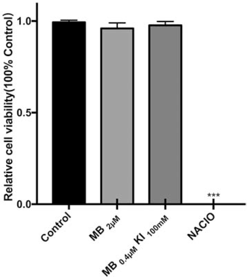
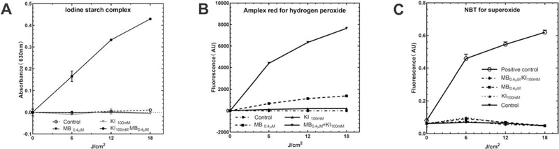
Results: KI could not only enhance MB aPDT on E. faecalis in both planktonic and biofilm states even in a hypoxic environment, but also produced a long-lasting bactericidal effect after end of the illumination. KI could accelerate photobleaching to reduce tooth staining by MB, and the mixture was harmless for HGFs. Mechanistic studies showed the generation of hydrogen peroxide and free iodine, and iodine radicals may be formed in hypoxia.
Conclusion: aPDT with MB plus KI could be used for root canal disinfection and clinical studies are worth pursuing.
Keywords: Antimicrobial photodynamic inactivation; Enterococcus faecalis; Methylene blue; Potassium iodide; Root canal disinfection.
Copyright © 2019 Elsevier B.V. All rights reserved.
Conflict of interest statement
Conflicts of interest
Figures
References
-
- Figdor D, Davies JK, Sundqvist G, Starvation survival, growth and recovery of Enterococcus faecalis in human serum, Oral Microbiol Immunol, 18 (2003) 234–239. - PubMed
-
- Lins RX, de Oliveira Andrade A, Hirata Junior R, Wilson MJ, Lewis MA, Williams DW, Fidel RA, Antimicrobial resistance and virulence traits of Enterococcus faecalis from primary endodontic infections, J Dent, 41 (2013) 779–786. - PubMed
MeSH terms
Substances
Grants and funding
LinkOut - more resources
Full Text Sources
Miscellaneous
