. 2020 Jan;34(1):8-17.
doi: 10.1097/BOT.0000000000001614.

Diagnosing Fracture-Related Infection: Current Concepts and Recommendations

Affiliations

Diagnosing Fracture-Related Infection: Current Concepts and Recommendations

Geertje A M Govaert et al. J Orthop Trauma. 2020 Jan.

Abstract

Fracture-related infection (FRI) is a severe complication after bone injury and can pose a serious diagnostic challenge. Overall, there is a limited amount of scientific evidence regarding diagnostic criteria for FRI. For this reason, the AO Foundation and the European Bone and Joint Infection Society proposed a consensus definition for FRI to standardize the diagnostic criteria and improve the quality of patient care and applicability of future studies regarding this condition. The aim of this article was to summarize the available evidence and provide recommendations for the diagnosis of FRI. For this purpose, the FRI consensus definition will be discussed together with a proposal for an update based on the available evidence relating to the diagnostic value of clinical parameters, serum inflammatory markers, imaging modalities, tissue and sonication fluid sampling, molecular biology techniques, and histopathological examination. Second, recommendations on microbiology specimen sampling and laboratory operating procedures relevant to FRI will be provided. LEVEL OF EVIDENCE:: Diagnostic Level V. See Instructions for Authors for a complete description of levels of evidence.


Conflict of interest statement

The authors report no conflict of interest.

Figures

FIGURE 1.
FIGURE 1.
Example of an x-ray (A) and T2-STIR MRI images (B coronal view, C sagittal view) performed in a patient with an infected proximal humerus fracture. The MRI demonstrated the extent of the infection with a skin defect, fluid/pus in the proximal humerus with surrounding edema, a sinus tract, and the involvement of the adjacent joint with possible involvement of the glenoid.
FIGURE 2.
FIGURE 2.
A 57-year-old male patient sustained a combined left-sided neck-of-femur fracture, a patella fracture, and a Gustilo-Anderson type IIIB open intra-articular distal femur fracture with a segmental defect due to a motor vehicle accident (A: x-ray left femur, AP). The open reduction and internal fixation was complicated by a FRI for which an induced membrane (Masquelet) procedure was carried out combined with a 3-month antibiotic treatment. Despite the fact that the patient was full–weight bearing and pain free, 2.5 years later, a fistula developed at his distal lateral left thigh. An x-ray (AP) of the left femur (B) showed that the implants were intact, the neck-of-femur fracture was healed and that there was bone formation on the medial side of the femoral fracture and over the lateral plate. There was no consolidation of the femoral defect. Preoperative workup included an FDG-PET/CT to assess the extent of the FRI. This scan demonstrated that the complete femur and all implants were involved in the infection and also visualized the trajectory of the soft tissue fistula. C–F, 18F-FDG PET/CT: (C) coronal FDG-PET image, (D) coronal fused FDG-PET/CT image, (E) sagittal fused FDG-PET/CT image, (F) transaxial FDG-PET/CT image with evidence of soft tissue fistula. (E) Preoperative clinical image with fistula (black arrow). (F) Intraoperative clinical image: The bone overlying the lateral plate is removed, and the infection revealed. AP, Antero-posterior.
FIGURE 3.
FIGURE 3.
A fracture-related infection, caused by Enterobacter cloacae, in a 48-year-old man following intramedullary nailing of the tibia. A, The patient presented with a draining fistula at the level of one of the distal locking screws, 4 months after placement of an intramedullary tibial nail. B, AP x-ray of the right lower leg showing the intramedullary nail of the tibia and plate osteosynthesis of the fibula. C–D, Delayed (4 hours) and late (20 hours) static time decay corrected planar images (anterior view) of the lower leg, showing focally increased accumulation of WBCs at the level of the fistula extending toward the tibia. Note the increased tracer accumulation and increase in extension over time, indicating the presence of an infection. E, A fused SPECT-CT image of WBC scintigraphy (4 hours after tracer injection) showing focally increased accumulation of WBCs at the level of the fistula extending around the distal part of the nail and the distal locking screw.
FIGURE 4.
FIGURE 4.
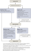
Descriptive flow chart of the diagnostic criteria of FRI. Adapted from Metsemakers et al, Injury 2018. Reprinted from Metsemakers WJ et al., Fracture-related infection: a consensus on definition from an international expert group. 2018 Injury:49/Issue 6, with permission from Elsevier. All permission requests for this image should be made to the copyright holder.

References

    1. Metsemakers WJ, Kortram K, Morgenstern M, et al. Definition of infection after fracture fixation: a systematic review of randomized controlled trials to evaluate current practice. Injury. 2018;49:497–504. - PubMed
    1. Metsemakers WJ, Moriarty TF, Morgenstern M, et al. Letter to the editor: new definition for periprosthetic joint infection: from the workgroup of the musculoskeletal infection Society. Clin Orthop Relat Res. 2016;474:2726–2727. - PMC - PubMed
    1. Parvizi J, Zmistowski B, Berbari EF, et al. New definition for periprosthetic joint infection: from the Workgroup of the Musculoskeletal Infection Society. Clin Orthop Relat Res. 2011;469:2992–2994. - PMC - PubMed
    1. Parvizi J, Tan TL, Goswami K, et al. The 2018 definition of periprosthetic hip and knee infection: an evidence-based and validated criteria. J Arthroplasty. 2018;33:1309–1314 e1302. - PubMed
    1. Morgenstern M, Moriarty TF, Kuehl R, et al. International survey among orthopaedic trauma surgeons: lack of a definition of fracture-related infection. Injury. 2018;49:491–496. - PubMed