Comparative Study
. 2019 Dec 19;14(12):e0225673.
doi: 10.1371/journal.pone.0225673. eCollection 2019.

4D perfusion CT of prostate cancer for image-guided radiotherapy planning: A proof of concept study

Affiliations
Comparative Study

4D perfusion CT of prostate cancer for image-guided radiotherapy planning: A proof of concept study

Lucian Beer et al. PLoS One. .

Abstract

Purpose: Advanced forms of prostate cancer (PCa) radiotherapy with either external beam therapy or brachytherapy delivery techniques aim for a focal boost and thus require accurate lesion localization and lesion segmentation for subsequent treatment planning. This study prospectively evaluated dynamic contrast-enhanced computed tomography (DCE-CT) for the detection of prostate cancer lesions in the peripheral zone (PZ) using qualitative and quantitative image analysis compared to multiparametric magnet resonance imaging (mpMRI) of the prostate.

Methods: With local ethics committee approval, 14 patients (mean age, 67 years; range, 57-78 years; PSA, mean 8.1 ng/ml; range, 3.5-26.0) underwent DCE-CT, as well as mpMRI of the prostate, including standard T2, diffusion-weighted imaging (DWI), and DCE-MRI sequences followed by transrectal in-bore MRI-guided prostate biopsy. Maximum intensity projections (MIP) and DCE-CT perfusion parameters (CTP) were compared between healthy and malignant tissue. Two radiologists independently rated image quality and the tumor lesion delineation quality of PCa using a five-point ordinal scale. MIP and CTP were compared using visual grading characteristics (VGC) and receiver operating characteristics (ROC)/area under the curve (AUC) analysis.

Results: The PCa detection rate ranged between 57 to 79% for the two readers for DCE-CT and was 92% for DCE-MRI. DCE-CT perfusion parameters in PCa tissue in the PZ were significantly different compared to regular prostate tissue and benign lesions. Image quality and lesion visibility were comparable between DCE-CT and DCE-MRI (VGC: AUC 0.612 and 0.651, p>0.05).

Conclusion: Our preliminary results suggest that it is feasible to use DCE-CT for identification and visualization, and subsequent segmentation for focal radiotherapy approaches to PCa.


Conflict of interest statement

The authors have declared that no competing interests exist.

Figures
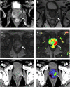
Fig 1
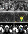
Fig 1. Examples of lesion delineation and image quality.
Images were obtained in a 78-year-old, biopsy-naive man. DWI images (A), 2D T2w images (B), dynamic contrast-enhanced MRI (C), pseudocolor maps (D), CT-MIP images (E), and CT images with blood flow overlay (F), in the axial plane are shown. In the pseudocolor map, tissue is colored according to its contrast uptake over time. Green areas represent a continuous uptake over time. Orange areas represent tissue that shows a plateau phase, and red areas represent tissue with a washout. A lesion was detected in the left peripheral zone (arrow). For lesion delineation and image quality, the score was excellent. Histopathology obtained by MRI-guided biopsy confirmed a PCa Gleason score 7 (3+4).
Fig 2
Fig 2. Examples of lesion delineation and image quality.
Images were obtained in a 63-year-old, biopsy-naive man. DWI images (A), 2D T2w images (B), dynamic contrast-enhanced MRI (C), pseudocolor maps (D), CT-MIP images (E), and CT images with blood flow overlay (F), in the axial plane are shown. In the pseudocolor map, tissue is colored according to its contrast uptake over time. Green areas represent a continuous uptake over time. Orange areas represent tissue that shows a plateau phase, and red areas represent tissue with a washout. A lesion was detected in the right peripheral zone (arrow). For lesion delineation and image quality, the score was acceptable. Histopathology obtained by MRI-guided biopsy confirmed a PCa Gleason score 7 (4+3).
Fig 3
Fig 3. Average time curves of contrast uptake in PCa versus healthy regular prostate tissue.
(A) Dynamic contrast-enhanced computed tomography (DCE-CT) and (B) dynamic contrast-enhanced magnetic resonance imaging (DCE-MRI), depicted for tumors in the peripheral zone. HU, Hounsfield Units.
Fig 4
Fig 4
Visual grading characteristic curves for lesion visibility (A, C) and image quality (B, D) assessment comparing DCE-CT and DCE-MRI (A, B) and DCE-CT MIP and CTP (C, D) reconstructions. R2 scored DCE-MRI significantly better than DCE-CT in terms of lesion visibility (VGC 0.73, p = 0.01) (A). There were no significant differences in terms of general image quality between DCE-CT and DCE-MRI (B). There was no significant difference between the different image reconstructions with regard to lesion visibility and image quality (p>0.05) (C, D). R, reader; VGC, visual grading characteristics; DCE-CT, dynamic contrast-enhanced computed tomography; DCE-MRI, dynamic contrast-enhanced magnet resonance imaging; MIP, maximum intensity projections; CTP, computed tomography perfusion parameters.

References

    1. Hollmann BG, van Triest B, Ghobadi G, Groenendaal G, de Jong J, et al. (2015) Gross tumor volume and clinical target volume in prostate cancer: How do satellites relate to the index lesion. Radiotherapy and oncology 115: 96–100. 10.1016/j.radonc.2015.01.021 - DOI - PubMed
    1. Daniel M, Kuess P, Andrzejewski P, Nyholm T, Helbich T, et al. (2018) Impact of androgen deprivation therapy on apparent diffusion coefficient and T2w MRI for histogram and texture analysis with respect to focal radiotherapy of prostate cancer. Strahlentherapie und Onkologie: 1–10. - PMC - PubMed
    1. Chopra S, Toi A, Taback N, Evans A, Haider MA, et al. (2012) Pathological predictors for site of local recurrence after radiotherapy for prostate cancer. International Journal of Radiation Oncology* Biology* Physics 82: e441–e448. - PubMed
    1. Monninkhof EM, van Loon JW, van Vulpen M, Kerkmeijer LG, Pos FJ, et al. (2018) Standard whole prostate gland radiotherapy with and without lesion boost in prostate cancer: Toxicity in the FLAME randomized controlled trial. Radiotherapy and Oncology 127: 74–80. 10.1016/j.radonc.2017.12.022 - DOI - PubMed
    1. Munck Af Rosenschold P, Zelefsky MJ, Apte AP, Jackson A, Oh JH, et al. (2018) Image-guided radiotherapy reduces the risk of under-dosing high-risk prostate cancer extra-capsular disease and improves biochemical control. Radiat Oncol 13: 64 10.1186/s13014-018-0978-1 - DOI - PMC - PubMed

Publication types

MeSH terms