

The prototype BS-II for computer measurement of biomechanical characteristics of the human cadaverous lumbar spine
- PMID: 31856862
- PMCID: PMC6924086
- DOI: 10.1186/s13018-019-1463-8
The prototype BS-II for computer measurement of biomechanical characteristics of the human cadaverous lumbar spine
Abstract
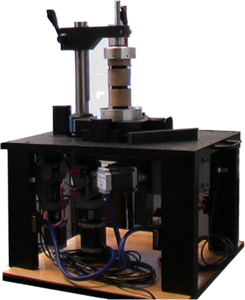
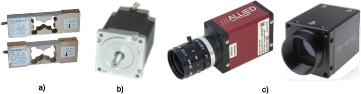
Background: The new second-generation computer system BS-II (Bio-Spine-II) based on the National Instruments' development environment has been designed and constructed for evaluating the stability of various surgical fixative methods of the cadaverous lumbar spine (L1-L5). BS-II holds the measured sample using aluminum fixtures and using four computer-controlled stepper motors; it performs a circular movement (warm up of the specimen), programmatically driven extension (back bend), right and left lateral flexion (lateral bend), left and right axial torsion (rotation), and axial compression (pressure). Four strain gauges are used to measure the stiffness of the sample. The movement of individual components (vertebrae) is contactlessly monitored by two CCD (charge couple device) cameras. The obtained data are in digital form continuously stored in the computer memory for further processing.
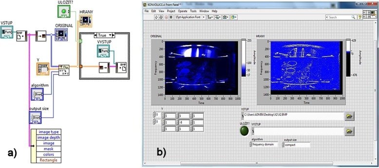
Methods: The functionality of the equipment was verified on the cadaverous specimen of the human spine. The stiffness of the sample was measured by strain gauges, and the results were processed using linear regression analysis. Movements of vertebrae were determined by circular discs covered with appropriate patterns. The discs have been linked with the respective vertebrae and were contactlessly monitored by two CCD (charge couple device) cameras and evaluated using digital image processing methods and 2D digital FFT (fast Fourier transformation). Direction and displacement of the individual components were determined by the band of the calculated spectrum. The new device BS-II is controlled by a modifiable computer program designed in the National Instruments' development environment which allows statistical processing of the sample, including its warming up.
Results: The computer system BS-II for measurement of biomechanical properties of the spine sample was designed. Functionality of the device was verified by implementation of LUMIR XLIF CAGE implant into a cadaver sample of the human spine. Comparison of the rigidity of the intact and stabilized sample is shown in the graphs of article. The achieved results contributed to certification of the implant into the surgical practice.
Conclusion: The designed computer BS-II system is designed for biomechanical measurements of the lumbar part of the human spine, especially for verification of surgical fixation methods. The system is based on the knowledge and experience with a manually operated measuring device designed by Palacky University Olomouc. The computer programmatic control allows the user to change the conditions and parameters of the measurement procedure in a planned way, which allows the results to be processed in, among other things, a statistical way. If suitable models are used (3D printing), the BS-II system can be used to verify procedures for surgical stabilization of the spine in the training of future doctors. The obtained data of stiffness and image information are stored in digital form and can be used for next offline sophisticated study of biomechanical properties of specimens (accurate vertebral geometry, statistical processing, 3D printing, etc.). The usefulness of the BS-II system is demonstrated in the case of biomechanical analysis of the implantation of LUMIR XLIF CAGE implant to a human cadaver specimen of the spine.
Keywords: BS-II; DIP; LabVIEW; Lumbar spine; Lumir XLIF CAGE; Strain gauge.
Conflict of interest statement
Not applicable.
Figures


References
MeSH terms
Grants and funding
LinkOut - more resources
Full Text Sources
