. 2019 Dec 19;50(1):110.
doi: 10.1186/s13567-019-0728-x.

Porcine rotavirus mainly infects primary porcine enterocytes at the basolateral surface

Affiliations

Porcine rotavirus mainly infects primary porcine enterocytes at the basolateral surface

Tingting Cui et al. Vet Res. .

Abstract

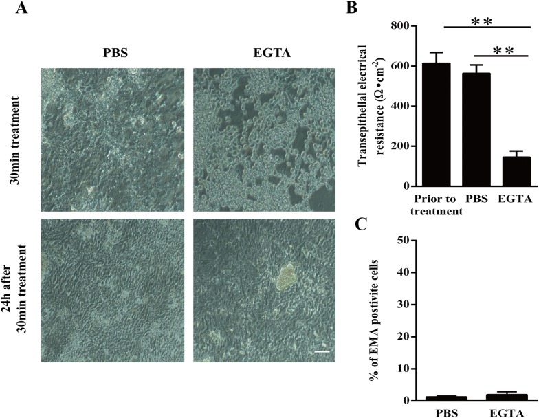
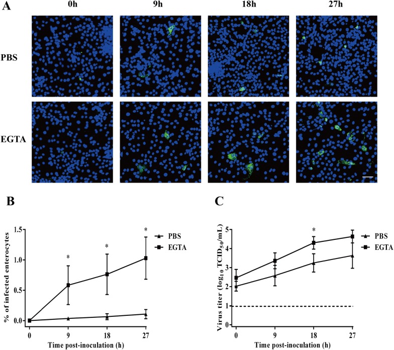
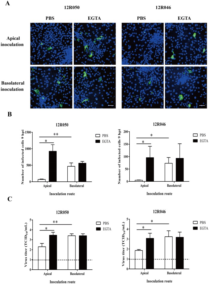
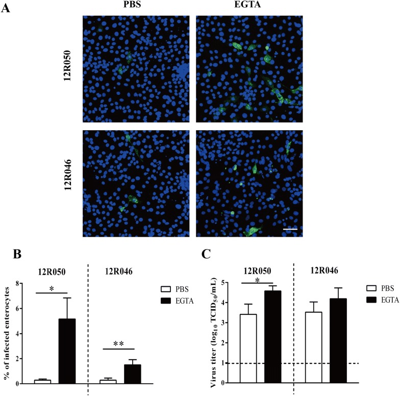
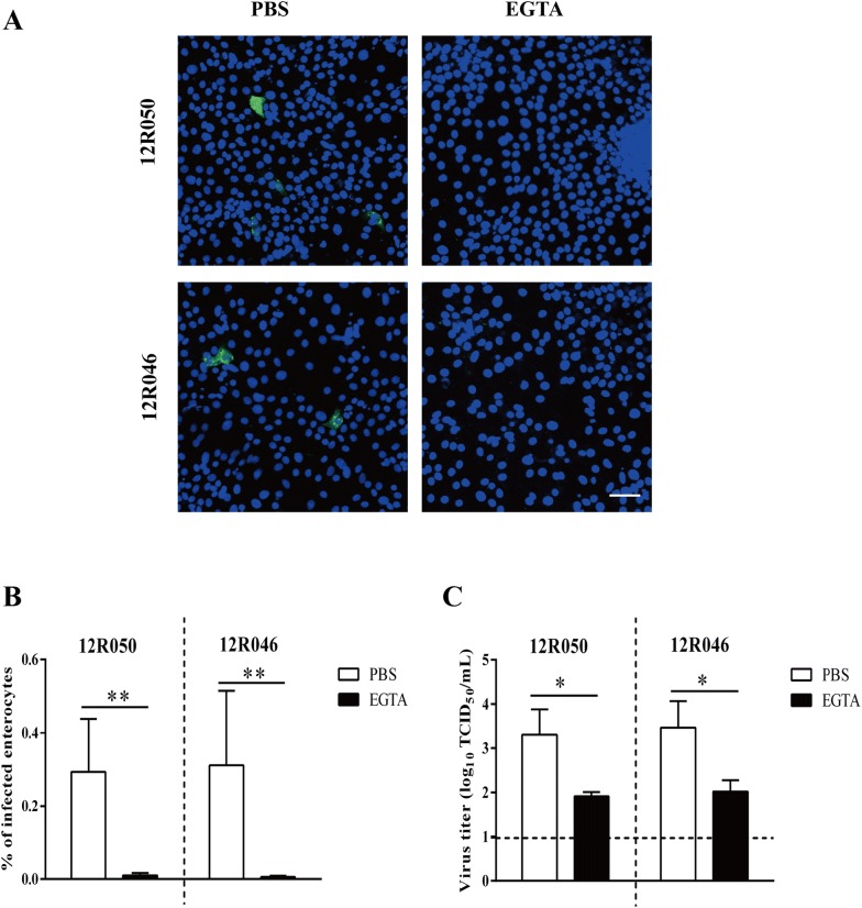
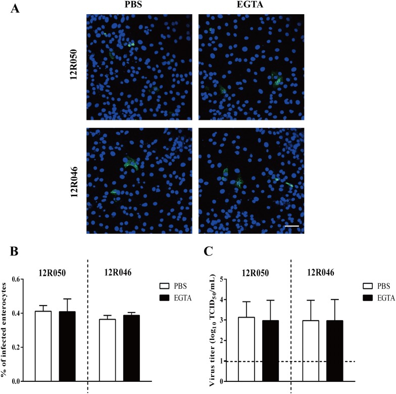
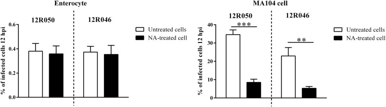
Intestinal epithelium functions as a barrier to protect multicellular organisms from the outside world. It consists of epithelial cells closely connected by intercellular junctions, selective gates which control paracellular diffusion of solutes, ions and macromolecules across the epithelium and keep out pathogens. Rotavirus is one of the major enteric viruses causing severe diarrhea in humans and animals. It specifically infects the enterocytes on villi of small intestines. The polarity of rotavirus replication in their target enterocytes and the role of intestinal epithelial integrity were examined in the present study. Treatment with EGTA, a drug that chelates calcium and disrupts the intercellular junctions, (i) significantly enhanced the infection of rotavirus in primary enterocytes, (ii) increased the binding of rotavirus to enterocytes, but (iii) considerably blocked internalization of rotavirus. After internalization, rotavirus was resistant to EGTA treatment. To investigate the polarity of rotavirus infection, the primary enterocytes were cultured in a transwell system and infected with rotavirus at either the apical or the basolateral surface. Rotavirus preferentially infected enterocytes at the basolateral surface. Restriction of infection through apical inoculation was overcome by EGTA treatment. Overall, our findings demonstrate that integrity of the intestinal epithelium is crucial in the host's innate defense against rotavirus infection. In addition, the intercellular receptor is located basolaterally and disruption of intercellular junctions facilitates the binding of rotavirus to their receptor at the basolateral surface.


Conflict of interest statement

The authors declare that they have no competing interests.

Figures

Figure 1
Figure 1
Disruption and restoration of intercellular junctions in primary enterocytes after EGTA treatment. A Representative microscopic images of enterocytes directly and 24 h after a 30 min treatment with EGTA. Scale bar: 100 µm. B Trans-epithelial electrical resistance of cells prior to treatment and after a 30-min treatment with PBS (control) or EGTA. C The percentage of EMA positive cells 24 h after treatment with PBS or EGTA. Data are expressed as the mean ± SD of the results of three separate experiments. Statistically significant (p < 0.01) differences are indicated with two asterisks.
Figure 2
Figure 2
Kinetics of rotavirus 12R050 (G5P[7]) replication in enterocytes after treatment with EGTA. A Representative confocal images of rotavirus infection (green) in primary enterocytes after a 30-min treatment with EGTA or PBS at 0, 9, 18 and 72 hpi. Scale bar: 50 µm. B The percentage of rotavirus infected enterocytes was analyzed by immunofluorescence staining. C Virus titer was determined in the supernatant with MA104 cells. Data are expressed as the mean ± SD of the results of three separate experiments. Statistically significant (p < 0.05) differences are indicated with an asterisk.
Figure 3
Figure 3
Rotavirus preferentially infects the basolateral surface of enterocytes and disruption of ICJ overcomes the restriction of rotavirus infection at the apical surface. A Representative confocal images of rotavirus infection (green) in enterocytes. Scale bar: 50 µm. To compare epithelial cell susceptibility to rotavirus, cells were exposed at either the apical surface or the basolateral surface to rotavirus 12R050 (G5P[7]) and 12R046 (G9P[23]). B The total number of infected cells per well was counted for each condition. C The virus titer was determined in supernatant for each condition. Data are expressed as the mean ± SD of the results of three separate experiments. Statistically significant differences are indicated with one asterisk (p < 0.05) and two asterisks (p < 0.01).
Figure 4
Figure 4
EGTA treatment increases the binding of rotavirus to enterocytes. Cells were pre-incubated with PBS or EGTA and inoculated with rotavirus at 4 °C for 1 h. After removing the unbound viral particles, cells were further incubated for 9 h. A Representative confocal image of infection in enterocytes. Scale bar: 50 µm. The percentage of infected cells (B) and the virus titer (C) were determined. Data are expressed as the mean ± SD of the results of three separate experiments. Statistically significant differences are indicated with one asterisk (p < 0.05) and two asterisks (p < 0.01).
Figure 5
Figure 5
EGTA treatment inhibits the internalization of rotavirus in enterocytes. Cells were inoculated with rotavirus at 4 °C for 1 h. After removing the unbound viral particles, cells were treated with EGTA or PBS at 37 °C for 30 min. Then, the cells were further incubated for 9 h after 3 washings with DMEM at 37 °C. A Representative confocal images of infection in enterocytes. Scale bar: 50 µm. The percentage of infected cells (B) and virus titer (C) were determined. Data are expressed as the mean ± SD of the results of three separate experiments. Statistically significant differences are indicated with one asterisk (p < 0.05) and two asterisks (p < 0.01).
Figure 6
Figure 6
EGTA treatment has no effect on the post-internalization stage of rotavirus particle in enterocytes. Cells were inoculated with rotavirus at 37 °C for 1 h. After 3 washings, cells were treated with EGTA or PBS for 30 min. Then, the cells were further incubated for 9 h at 37 °C. A Representative confocal image of infection in enterocytes. Scale bar: 50 µm. The percentage of infected cells (B) and virus titer (C) were determined. Data are expressed as the mean ± SD of the results of three separate experiment.
Figure 7
Figure 7
Effect of neuraminidase treatment on the replication of rotaviruses in primary enterocytes and MA104 cells. Primary enterocytes and MA104 Cells were treated with 50 mU/mL NA from Vibrio Cholerae for 1 h at 37 °C before inoculation with rotavirus at a MOI of 0.1. The percentage of infection was measured by immunofluorescence staining at 12 hpi. Data are expressed as the mean ± SD of the results of three separate experiments. Statistically significant differences are indicated with two asterisks (p < 0.01) and three asterisks (p < 0.001).

Similar articles

Cited by

References

    1. Trejo-Cerro Ó, Eichwald C, Schraner EM, Silva-Ayala D, López S, Arias CF. Actin-dependent nonlytic rotavirus exit and infectious virus morphogenetic pathway in nonpolarized cells. J Virol. 2018;92:e02076-17. doi: 10.1128/JVI.02076-17. - DOI - PMC - PubMed
    1. Estes MK, Graham D, Gerba CP, Smith E. Simian rotavirus SA11 replication in cell cultures. J Virol. 1979;31:810–815. - PMC - PubMed
    1. Guerrero CA, Zarate S, Corkidi G, López S, Arias CF. Biochemical characterization of rotavirus receptors in MA104 cells. J Virol. 2000;74:9362–9371. doi: 10.1128/JVI.74.20.9362-9371.2000. - DOI - PMC - PubMed
    1. Liu F, Li G, Wen K, Bui T, Cao D, Zhang Y, Yuan L. Porcine small intestinal epithelial cell line (IPEC-J2) of rotavirus infection as a new model for the study of innate immune responses to rotaviruses and probiotics. Viral Immunol. 2010;23:135–149. doi: 10.1089/vim.2009.0088. - DOI - PMC - PubMed
    1. Obert G, Peiffer I, Servin AL. Rotavirus-induced structural and functional alterations in tight junctions of polarized intestinal Caco-2 cell monolayers. J Virol. 2000;74:4645–4651. doi: 10.1128/JVI.74.10.4645-4651.2000. - DOI - PMC - PubMed

LinkOut - more resources