. 2020 Jan 15;26(2):450-464.
doi: 10.1158/1078-0432.CCR-19-2309. Epub 2019 Dec 19.

Mutation-derived Neoantigen-specific T-cell Responses in Multiple Myeloma

Affiliations

Mutation-derived Neoantigen-specific T-cell Responses in Multiple Myeloma

Deepak Perumal et al. Clin Cancer Res. .

Abstract

Purpose: Somatic mutations in cancer cells can give rise to novel protein sequences that can be presented by antigen-presenting cells as neoantigens to the host immune system. Tumor neoantigens represent excellent targets for immunotherapy, due to their specific expression in cancer tissue. Despite the widespread use of immunomodulatory drugs and immunotherapies that recharge T and NK cells, there has been no direct evidence that neoantigen-specific T-cell responses are elicited in multiple myeloma.

Experimental design: Using next-generation sequencing data we describe the landscape of neo-antigens in 184 patients with multiple myeloma and successfully validate neoantigen-specific T cells in patients with multiple myeloma and support the feasibility of neoantigen-based therapeutic vaccines for use in cancers with intermediate mutational loads such as multiple myeloma.

Results: In this study, we demonstrate an increase in neoantigen load in relapsed patients with multiple myeloma as compared with newly diagnosed patients with multiple myeloma. Moreover, we identify shared neoantigens across multiple patients in three multiple myeloma oncogenic driver genes (KRAS, NRAS, and IRF4). Next, we validate neoantigen T-cell response and clonal expansion in correlation with clinical response in relapsed patients with multiple myeloma. This is the first study to experimentally validate the immunogenicity of predicted neoantigens from next-generation sequencing in relapsed patients with multiple myeloma.

Conclusions: Our findings demonstrate that somatic mutations in multiple myeloma can be immunogenic and induce neoantigen-specific T-cell activation that is associated with antitumor activity in vitro and clinical response in vivo. Our results provide the foundation for using neoantigen targeting strategies such as peptide vaccines in future trials for patients with multiple myeloma.


Figures

Figure 1.
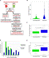
Figure 1.. High frequency of neoantigens observed in relapsed myeloma patients as compared to newly diagnosed MM patients
A, Neoantigen discovery pipeline used in this study. B, Distribution of mutational burden (ie, number of somatic mutations per megabase [Mb] detected in newly diagnosed and relapsed MM patients from whole-exome sequencing (WES) data. C, High mutational load in relapsed MM patients as compared to newly diagnosed myeloma patients (p <0.0001, Wilcoxon Rank Sum Test). D, Frequency of predicted neoantigens in newly diagnosed and relapsed MM patients. E, High frequency of neoantigen load in relapsed MM patients as compared to newly diagnosed myeloma patients (p <0.0001, Wilcoxon Rank Sum Test).
Figure 2.
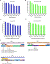
Figure 2.. Neoantigens are observed in recurrently mutated MM genes
A, The top 10 recurrent somatic mutations observed in 92 relapsed MM patients. NRASQ61R was the top recurrent mutation in our relapsed MM patients, B, The top 10 recurrent somatic mutations observed in 92 newly diagnosed MM patients. CDC27L85F was the top recurrent mutation in our newly diagnosed MM patients. C, The top 10 most frequently observed immunogenic mutations in relapsed MM patients. Mutated genes that could yield potentially immunogenic neoantigens in relapsed MM patients were PKD1, EP400, PRKDC, NRAS, UBR4, KIAA2013, ATM, MED12, IPO7 and MYCPB2. For the majority of the identified neo-antigens, we observed that mutations in the same gene are not shared between relapsed MM patients and are highly patient-specific D, The top 10 most frequently observed immunogenic mutations in newly diagnosed MM patients. In newly diagnosed MM patients LAMA5, DYNC1H1, FASN, UBR4, NCOR2, RNF213, KRAS, UBR5, VPS13A and PRKDC were identified. For the majority of the identified neo-antigens, we observed that mutations in the same gene are not shared between newly diagnosed MM patients and are highly patient-specific E, Shared neoantigens identified in MM patients were found in recurrently mutated genes KRAS, NRAS and IRF4. Shared neoantigens in KRAS, NRAS and IRF4 and their corresponding mutants in observed in both relapsed and newly diagnosed MM patients are shown in the plot. We observed that 5 relapsed patients shared NRASQ61R mutations (Strong binding, IC50 < 150 nM), 4 relapsed patients shared IRF4K123R mutations (Very strong binding, IC50 < 50 nM), 3 relapsed patients shared KRASQ61H mutations (Strong binding, IC50 < 150 nM) and 2 relapsed patients shared NRASQ61K mutations (Strong binding, IC50 < 150 nM) (Fig. 2c). Similarly, we observed that 3 newly diagnosed patients shared the KRASQ61H mutations (Strong binding, IC50 < 150 nM), 2 newly diagnosed patients shared KRASG12V neoantigenic mutations (Very strong binding, IC50 < 50 nM) and 2 newly diagnosed patients shared KRASQ61R neoantigenic mutations (Intermediate binding, IC50 of 150–250 nM).
Figure 3.
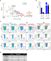
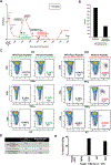
Figure 3.. Checkpoint based inhibitor therapy elicits PRKDC-neoantigen specific T cell response in a primary refractory MM patient
A, Timeline of clinical response of MM patient to dual checkpoint inhibitor (anti-CTLA4 +anti-PD-1) therapy. B, The number of non-synonymous mutations and the predicted immunogenic neoantigens. C, CD8+ T cell response to neoantigen peptide (PRKDC) measured by IFNg, TNFa and IL2 pre & post dual checkpoint inhibition (anti-CTLA4 + anti-PD-1). D, The clinical response and T cell response to neoantigen of this patient prior and post to immunotransplant and checkpoint inhibition.
Figure 4.
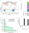
Figure 4.. Assessment of CD8+ T Cell Responses to PRKDC neoantigen using MHC Class I Tetramer, TCR sequencing and Cytotoxic T cell killing
A, Validation of HLA-A*03:01 tetramer with neoantigen peptide PRKDC from the MM patient. Peripheral lymphocytes obtained post checkpoint blockade treatment was incubated with pMHC tetramer (HLA-A03:01). There was a significant increase in tetramer positive CD8+ T cells with the mutant peptide (1.51%) as compared to the wild type peptide (0.3%). B, TCR-Seq reveals increase in oligoclonal expansion after dual checkpoint inhibition. Tetramer-binding T cells were subjected for sequencing of T-cell receptor (TCR) β-chains. We observed peripheral T-cell expansion of the top 10 most dominant intratumoral clones, with the most dominant clones reaching a 1.464 % and 1.152 % increase in abundance in the ex vivo expanded cells post immune checkpoint therapy than at the time of pretreatment (cells without ex vivo expansion). C, The specific TCR-β clones are shown in the bar plot. The patient had a high proportion of pre-existing dominant clones after the administration of checkpoint therapy compared to the low proportion of such pre-existing dominant clones before ASCT and checkpoint therapy. D, Neoantigen Specific CD8+ T cell Cytotoxicity. Antigen specific effector T cells were expanded with peptides (Mutant (Mut), Wild Type (WT), CEFT (+ ctrl) and MOG (-ve ctrl) for 10 days following which patients target (T) cells were incubated with expanded effector (E) T cells with T:E ratio of 1:100. Shown is the antigen specific lysis in % which is the specific lysis specific to appropriate peptides.
Figure 5.
Figure 5.. Checkpoint blockade therapy enhances EVI2B-neoantigen specific T cell response in a relapsed MM patient
A, Timeline of clinical response of MM patient to dual checkpoint inhibitor (anti-CTLA4 +anti-PD-1) therapy. B, The number of non-synonymous mutations and the predicted immunogenic neoantigens. C, CD8+ T cell response to neoantigen peptide (EVI2B) measured by IFNg, TNFa and IL2 pre & post dual checkpoint inhibition (anti-CTLA4 + anti-PD-1). D, The clinical response and T cell response to neoantigen of this patient prior and post to immunotransplant and checkpoint inhibition. E, Neoantigen Specific CD8+ T cell Cytotoxicity. Antigen specific effector T cells were expanded with peptides (Mutant (Mut), Wild Type (WT), CEFT (+ ctrl) and MOG (-ve ctrl) for 10 days following which patients target (T) cells were incubated with expanded effector (E) T cells with T:E ratio of 1:100. Shown is the antigen specific lysis in % which is the specific lysis specific to appropriate peptides.
Figure 6.
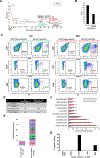
Figure 6.. Checkpoint blockade therapy in combination with Pomalidomide and elotuzumab treatment elicits S100A9-neoantigen specific T cell response in a relapsed MM patient
A, Timeline of clinical response of MM patient to checkpoint inhibitor anti-PD-L1 + elotuzumab and pomalidomide therapies. B, The number of non-synonymous mutations and the predicted immunogenic neoantigens. C, CD8+ T cell response to neoantigen peptide (S100A9) measured by IFNg, TNFa and IL2 pre & post dual treatment (anti-PD-L1 + elotuzumab and pomalidomide). D, The clinical response and T cell response to neoantigen of this patient prior and post to checkpoint inhibition and elotuzumab plus pomalidomide treatments. E, TCR-Seq reveals increase in oligoclonal expansion after anti-PD-L1 + elotuzumab and pomalidomide treatments. We observed peripheral T-cell expansion of a subset of the top 10 most dominant intratumoral clones, with the most dominant clones reaching a 1.261 % and 0.996 % increase in abundance in the blood post treatment (without ex vivo expansion). F, The specific TCR-β clones are shown in the bar plot. The patient had a high proportion of pre-existing dominant clones after the administration of immunomodulatory therapies compared to the low proportion of such pre-existing dominant clones before treatment. G, Neoantigen Specific CD8+ T cell Cytotoxicity. Antigen specific effector T cells were expanded with peptides (Mutant (Mut), Wild Type (WT), CEFT (+ ctrl) and MOG (-ve ctrl) for 10 days following which patients target (T) cells were incubated with expanded effector (E) T cells with T:E ratio of 1:100. Shown is the antigen specific lysis in % which is the specific lysis specific to appropriate peptides.

References

    1. Kuehl WM, Bergsagel PL. Molecular pathogenesis of multiple myeloma and its premalignant precursor. The Journal of Clinical Investigation 2012;122(10):3456–63. - PMC - PubMed
    1. Siegel RL, Miller KD, Jemal A. Cancer statistics, 2016. CA: A Cancer Journal for Clinicians 2016;66(1):7–30. - PubMed
    1. Rajasagi M, Shukla SA, Fritsch EF, Keskin DB, DeLuca D, Carmona E, et al. Systematic identification of personal tumor-specific neoantigens in chronic lymphocytic leukemia. Blood 2014;124(3):453. - PMC - PubMed
    1. Rizvi NA, Hellmann MD, Snyder A, Kvistborg P, Makarov V, Havel JJ, et al. Mutational landscape determines sensitivity to PD-1 blockade in non–small cell lung cancer. Science (New York, NY) 2015;348(6230):124–8. - PMC - PubMed
    1. Snyder A, Makarov V, Merghoub T, Yuan J, Zaretsky JM, Desrichard A, et al. Genetic Basis for Clinical Response to CTLA-4 Blockade in Melanoma. New England Journal of Medicine 2014;371(23):2189–99. - PMC - PubMed

Publication types

MeSH terms