. 2020 Jan 14;52(1):123-135.e6.
doi: 10.1016/j.immuni.2019.11.013. Epub 2019 Dec 16.

Immune Sensing of Cell Death through Recognition of Histone Sequences by C-Type Lectin-Receptor-2d Causes Inflammation and Tissue Injury

Affiliations

Immune Sensing of Cell Death through Recognition of Histone Sequences by C-Type Lectin-Receptor-2d Causes Inflammation and Tissue Injury

Jiann-Jyh Lai et al. Immunity. .

Abstract

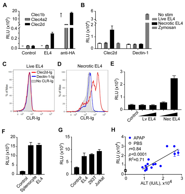
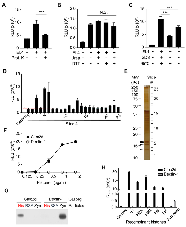
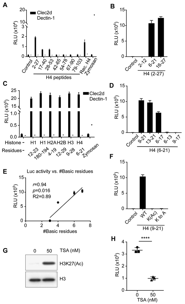
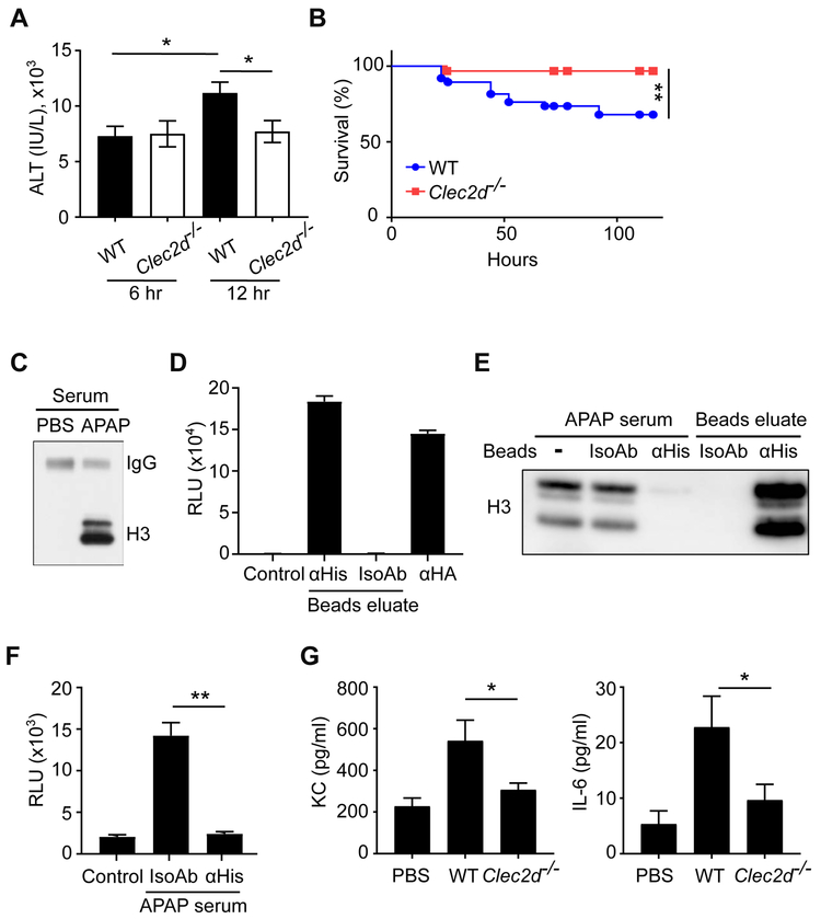
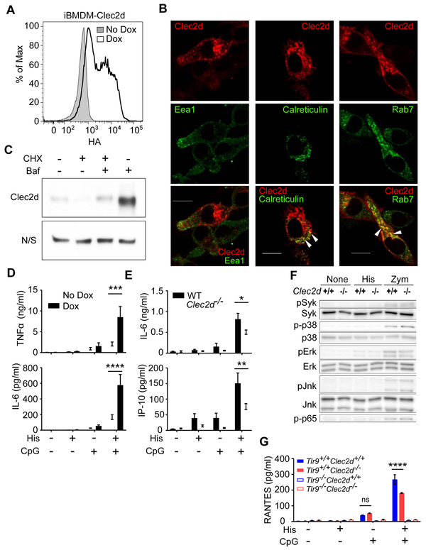
The immune system monitors the health of cells and is stimulated by necrosis. Here we examined the receptors and ligands driving this response. In a targeted screen of C-type lectin receptors, a Clec2d reporter responded to lysates from necrotic cells. Biochemical purification identified histones, both free and bound to nucleosomes or neutrophil extracellular traps, as Clec2d ligands. Clec2d recognized poly-basic sequences in histone tails and this recognition was sensitive to post-translational modifications of these sequences. As compared with WT mice, Clec2d-/- mice exhibited reduced proinflammatory responses to injected histones, and less tissue damage and improved survival in a hepatotoxic injury model. In macrophages, Clec2d localized to the plasma membrane and endosomes. Histone binding to Clec2d did not stimulate kinase activation or cytokine production. Rather, histone-bound DNA stimulated endosomal Tlr9-dependent responses in a Clec2d-dependent manner. Thus, Clec2d binds to histones released upon necrotic cell death, with functional consequences to inflammation and tissue damage.

Keywords: C-type lectin; Clec2d; DAMPs; Toll-like receptor; histone acetylation; histones; inflammation; liver injury; macrophages; pattern recognition receptor.


Conflict of interest statement

DECLARATION OF INTERESTS

The authors declare no competing interests.

Figures

Figure 1.
Figure 1.. Using a Reporter System to Screen for Novel DAMP Receptors.
(A) RF33.70-Luc reporters expressing the indicated CLR-CD3ζ chimeric receptors were stimulated overnight with or without necrotic EL4 cell lysates, and then luciferase activity was assayed. Anti-HA (αHA) was used to stimulate reporter cells as a positive control. Control, reporter cells in media without necrotic lysates. (B) Clec2d or Dectin-1 reporters were left without stimulation (No stim) or stimulated with supernatant from live or necrotic EL4 cultures, and the luciferase activity was measured. Zymosan was used to stimulate Dectin-1 reporters as a positive control. (C, D) Live or necrotic EL4 cells were incubated with soluble Clec2d-Ig or Dectin-1-Ig, followed by staining with secondary antibodies conjugated with fluorophore and analyzed by flow cytometry. (E) Live or necrotic EL4 cells (1x10⁵ or 3x10⁵/well) were fixed with paraformaldehyde and then used to stimulate Clec2d reporters, and the luciferase activity was measured. (F) Reporter cells expressing Clec2d chimeric receptors were stimulated without (control) or with necrotic mouse splenocytes or EL4 cells overnight, and then luciferase activity was measured. (G) Clec2d reporter cells were stimulated overnight without (control) or with necrotic EL4, 293T, or Jurkat cells and then luciferase activity was measured. (H) Six or 22 hr after APAP (300 or 500 mg/KgBW) or PBS injection into WT mice, serum was collected and assayed for ALT activity and stimulation of Clec2d reporter cells. Pearson correlation coefficient analysis of Clec2d reporter activity versus serum ALT activity, with r=0.84, p<0.0001, R2=0.71. See also Figure S1.
Figure 2.
Figure 2.. Characterization of the Necrotic Cell Ligands Recognized by Clec2d.
(A) Necrotic EL4 lysates were treated without or with Proteinase K-agarose beads (0.35 unit/ml) at 37°C for 4 hr. The protease beads were then removed and the lysates were tested for their ability to stimulate Clec2d reporter cells to produce luciferase. ***, p<0.001 with One-way ANOVA. (B) Necrotic EL4 lysates were treated with or without 8 M urea and/or 20 mM DTT at RT for 30 min, and then assayed for their ability to stimulate Clec2d reporter cells to produce luciferase. N.S., no significant difference. (C) Necrotic EL4 lysates were treated with or without 2% SDS and/or heated at 95°C for 10 min. These lysates were then used to stimulated Clec2d reporter cells to produce luciferase. ***, p<0.001 with One-way ANOVA. (D, E) The subcellular fraction enriched with ligand activity (See also Figure S2.) was further fractionated by SDS-PAGE. Part of the gel was used for silver stain to reveal protein bands (E), and the rest of the gel was transferred onto PVDF membrane, and sliced into 23 fractions. Each membrane slice was co-cultured with Clec2d reporter cells, and then luciferase activity was measured (D). Protein bands (arrow heads) in gel fractions with the highest luciferase activity (#3, 5 and 6) were analyzed by Mass Spectrometry, which revealed the presence of histone H4, H2A/H2B, and H3 as the major proteins in each fraction, respectively. (F) Reporter cells with Clec2d or Dectin-1 chimeric receptors were stimulated with purified histones from calf thymus at the indicated concentrations. Luciferase activity was measured in the lysate after overnight stimulation. (G) Zymosan particles (Zym) or agarose beads conjugated with histones (His) or BSA (BSA) were incubated with soluble Clec2d or Dectin-1 receptors expressed as mouse IgG Fc fusion proteins. Proteins that bound these particles were eluted, and analyzed with anti-mouse IgG antibodies by western blots. (H) Reporter cells with Clec2d or Dectin-1 chimera receptors were stimulated overnight with the indicated recombinant human histone proteins or zymosan and then luciferase activity was measured. Control = reporter cells cultured in media without histones or zymosan. See also Figure S2 and S3.
Figure 3.
Figure 3.. Mapping the Clec2d-Binding Regions of Histone H4.
(A) Overlapping synthetic peptides of mouse histone H4 or no peptide (control) were tested for their ability to stimulate Clec2d or Dectin-1 reporter cells to produce luciferase activity. Zymosan and recombinant H4 (Rec. H4) were used as positive controls. The H4 number ranges denote the amino acid positions within the H4 sequence of the synthetic peptides, where 1 is the N-terminal residue). (B) Smaller synthetic peptides located at the N-terminal (H42-12), middle (H46-21), or C-terminal (H416-27) of H42-27 were used to stimulate Clec2d reporters and the luciferase activity was measured. Control = reporter cells cultured without peptides. (C) Similar to (B), synthetic peptides of the tail regions in all 5 histones were used to stimulate the Clec2d reporters. Zymosan was used as a positive control for stimulation of the Dectin-1 reporters. (D) Similar to (B) but the smaller synthetic peptides were within H46-21 containing different numbers of basic residues. Control = reporter cells cultured without peptides. (E) Pearson correlation coefficient analysis of (D) of Clec2d reporter activity versus number of basic residues in the synthetic peptides, with r=0.94, p=0.016, R2=0.89. (F) Similar to (B) except the peptides were H49-21 or its derivative peptides, “K(Ac)” peptide with all lysine residues acetylated, or “K to A” peptide with all lysine residues replaced by alanine. Control = reporter cells cultured without peptides. (G) EL4 cells were treated with or without TSA (50 nM) for 19 hr and then disrupted by freeze and thaw cycles. The resulting lysates were analyzed by western blot and probed for H3K27 acetylation or total H3 histone, or (H) used to stimulate Clec2d reporter cells and after which luciferase activity was measured. ****, p<0.0001, with Student’s t-test. See also Figure S4.
Figure 4.
Figure 4.. Clec2d Enhanced Liver Injury in Acetaminophen Overdose
(A) WT and Clec2d−/− mice were fasted for 24 hr and then injected i.p. with 300 mg/KgBW APAP. Six or 12 hr after APAP injection, serum ALT activity was measured. 6 hr (WT), n=26; 6 hr (Clec2d−/−), n=17; 12 hr (WT), n=54; 12 hr (Clec2d−/−) n=42. Mean ± SEM. *, p<0.05 by One-Way ANOVA. (B) Survival of the mice was monitored for 5 days after injection. WT, n=38; Clec2d−/−, n=32. Survival curve comparison was analyzed by a Log-rank Mantel-Cox test. **, p<0.01. (C) Western blot to detect histone H3 in the serum of WT mice with or without APAP treatment. Mouse serum IgG was also detected on the same blot as a loading control. (D) Histones from the serum of APAP-treated mice were purified by anti-histone (αHis) or isotype control (IsoAb) antibody-conjugated beads. The eluate from the beads were immobilized on the assay plate to stimulate reporter cells expressing Clec2d and luciferase activity was measured after 20 hr. Anti-HA (αHA) antibody was used as a positive control for reporter stimulation. Control=reporter cells in media. (E, F) Histones in the serum of APAP-treated mice (APAP serum) were immunodepleted by anti-histone (αHis) or isotype control (IsoAb) antibody-conjugated beads. Histone H3 in the post-treatment serum or bead-eluates was detected by western blot (E). The APAP serum after treatment was immobilized on a plate and used to stimulate the Clec2d reporter cells (F). Control = reporter cells in media. Mean ± SEM. **, p<0.01 by One-Way ANOVA. (G) WT and Clec2d−/− mice were i.v. injected with purified histones, and 18 hr later cytokine and chemokine concentrations were measured in the serum. WT and Clec2d−/−, n=7; PBS control, n=5. Mean ± SEM. *, p<0.05 by One-Way ANOVA.
Figure 5.
Figure 5.. Characterization of Clec2d in Macrophages
(A) iBMDM cells transduced with a Dox-inducible Clec2d-HA construct were cultured with or without Dox and then analyzed for surface HA staining by flow cytometry. (B) iBMDM cells transfected with a Dox-inducible Clec2d-mCherry construct (Red) were cultured with Dox, permeabilized, stained (green) for Eea1, Rab7, or Calreticulin, and analyzed by confocal microscopy. Colocalization of Clec2d with Rab7 or calreticulin is shown in yellow in the merged images and highlighted with arrows. Bars=10 μm (C) Western blot data showing that Bafilomycin (Baf) rescues total Clec2d amounts when peritoneal macrophages were treated with Cycloheximide (CHX). N/S = non-specific band on the same αClec2d blot, which served as a loading control. (D) iBMDM cells transduced with a Dox-inducible Clec2d construct were treated with or without Dox and then stimulated for overnight with or without histones (30 μg/ml) and/or CpG (1 μg/ml). TNFα and IL-6 were measured in the culture supernatant by ELISA. The data was pooled from 5 experiments. Mean ± SEM. ***, p<0.001; ****, p<0.0001 by Two-Way ANOVA. (E) Primary peritoneal macrophages were stimulated for overnight with 30 μg/ml histones and/or 0.1 μg/ml CpG. IL-6 and IP-10 were measured in the supernatant. Mean ± SEM, *, p<0.05; **, p<0.01 by Two-Way ANOVA. (F) Peritoneal macrophages from WT and Clec2d−/− mice were plated and stimulated without (None) or with 30 μg/ml histones (His) or 40 μg/ml zymosan (Zym) for 30 min. The phosphorylation of MAPKs, NFκB (p65), and Syk were detected in the cell lysates by western blot. (G) Peritoneal macrophages from WT, Clec2d−/− and/or Tlr9−/− mice were stimulated for overnight with or without histones (30 μg/ml) and/or CpG (0.1 μg/ml). RANTES was measured in the supernatant. Mean ± SEM, ****, p<0.0001 by Two-Way ANOVA. ns, not significant. See also Figure S5.
Figure 6.
Figure 6.. Clec2d Recognizes Physiological Ligands
(A) Purified nucleosomes or histones were coated on a plate, cultured with reporter cells expressing Clec2d, and luciferase activity was measured. (B) Peritoneal macrophages from WT or Clec2d−/− mice were stimulated with purified nucleosomes for 18 hr and the RANTES concentration in the supernatant was measured by ELISA. (C) Purified nucleosomes were pre-treated with or without DNase I at 37°C for 1 hr and used to stimulate peritoneal macrophages from WT and Clec2d−/− mice. RANTES concentration was measured by ELISA in the supernatant after 18 hr stimulation. DNase I alone serves as a control. Mean ± SEM , ****, P<0.001 by Two-Way ANOVA. (D) Neutrophils isolated from WT mice were treated with 10 μM Ionomycin for 4 hr to induce NETs formation in a 96-well plate. Reporter cells expressing Clec2d or Dectin-1 were then cultured in the plate and luciferase activity was measured after 20 hr stimulation. Immobilized histones (for Clec2d reporters) and zymosan (for Dectin-1 reporters) serve as positive controls for reporter stimulation. (E) Purified neutrophils were treated with 10 μM Ionomycin for 4 hr to induce NETs formation in the chamber slide. After treatment with 2% paraformaldehyde for 5 min, the NETs were stained with Clec2d-Ig or Dectin-1-Ig followed by secondary antibodies conjugated with fluorophore. The slides were then mounted with mounting oil containing DAPI. Bars=20 μm.

Comment in

References

    1. Ahrens S, Zelenay S, Sancho D, Hanc P, Kjaer S, Feest C, Fletcher G, Durkin C, Postigo A, Skehel M, et al. (2012). F-actin is an evolutionarily conserved damage-associated molecular pattern recognized by DNGR-1, a receptor for dead cells. Immunity 36, 635–645. - PubMed
    1. Alcover A, Alarcon B, and Di Bartolo V (2018). Cell Biology of T Cell Receptor Expression and Regulation. Annu Rev Immunol 36, 103–125. - PubMed
    1. Allam R, Kumar SV, Darisipudi MN, and Anders HJ (2014). Extracellular histones in tissue injury and inflammation. J Mol Med (Berl) 92, 465–472. - PubMed
    1. Allam R, Scherbaum CR, Darisipudi MN, Mulay SR, Hagele H, Lichtnekert J, Hagemann JH, Rupanagudi KV, Ryu M, Schwarzenberger C et al. (2012). Histones from dying renal cells aggravate kidney injury via TLR2 and TLR4. J Am Soc Nephrol 23, 1375–1388. - PMC - PubMed
    1. Augusto LA, Decottignies P, Synguelakis M, Nicaise M, Le Marechal P, and Chaby R (2003). Histones: a novel class of lipopolysaccharide-binding molecules. Biochemistry 42, 3929–3938. - PubMed

Publication types

MeSH terms