. 2019 Dec 20;14(12):e0226260.
doi: 10.1371/journal.pone.0226260. eCollection 2019.

In vitro activity and In vivo efficacy of Isoliquiritigenin against Staphylococcus xylosus ATCC 700404 by IGPD target

Affiliations

In vitro activity and In vivo efficacy of Isoliquiritigenin against Staphylococcus xylosus ATCC 700404 by IGPD target

Qianwei Qu et al. PLoS One. .

Abstract

Staphylococcus xylosus (S. xylosus) is a type of coagulase-negative Staphylococcus, which was previously considered as non-pathogenic. However, recent studies have linked it with cases of mastitis in cows. Isoliquiritigenin (ISL) is a bioactive compound with pharmacological functions including antibacterial activity. In this study, we evaluated the effect of ISL on S. xylosus in vitro and in vivo. The MIC of ISL against S. xylosus was 80 μg/mL. It was observed that sub-MICs of ISL (1/2MIC, 1/4MIC, 1/8MIC) significantly inhibited the formation of S. xylosus biofilm in vitro. Previous studies have observed that inhibiting imidazole glycerol phosphate dehydratase (IGPD) concomitantly inhibited biofilm formation in S. xylosus. So, we designed experiments to target the formation of IGPD or inhibits its activities in S. xylosus ATCC 700404. The results indicated that the activity of IGPD and its histidine content decreased significantly under 1/2 MIC (40 μg/mL) ISL, and the expression of IGPD gene (hisB) and IGPD protein was significantly down-regulated. Furthermore, Bio-layer interferometry experiments showed that ISL directly interacted with IGPD protein (with strong affinity; KD = 234 μM). In addition, molecular docking was used to predict the binding mode of ISL and IGPD. In vivo tests revealed that, ISL significantly reduced TNF-α and IL-6 levels, mitigated the destruction of the mammary glands and reversed the production of inflammatory cells in mice. The results of the study suggest that, ISL may inhibit S. xylosus growth by acting on IGPD, which can be used as a target protein to treat infections caused by S. xylosus.


Conflict of interest statement

The authors have declared that no competing interests exist.

Figures

Fig 1
Fig 1. The chemical structure of isoliquiritigenin.
Fig 2
Fig 2. Time-kill curves of ISL against S. xylosus cells.
Fig 3
Fig 3
Determination of biofilm formation ability (A and B) and scanning electron microscope observation (C, D, E and F) of S. xylosus (wild or mutant). (A) Effect of sub-MICs of ISL on biofilm formation by wild-type S. xylosus strain. Data are expressed as means ± SDs. Controls refer to the absence of ISL. Significantly different means are indicated with asterisks (*) (*p < 0.05, **p < 0.01, and ***p < 0.001) compared to the untreated control group. (B) Effect of sub-MICs of ISL on biofilm formation by mutant S. xylosus ATCC700404 strain. Data are expressed as means ± SDs. Controls refer to the absence of ISL. Means without asterisks (*) have no significant difference compared to the untreated control group. (C) Untreated wild-type strains; (D) Wild-type strains treated with 1/2 MIC of ISL (40μg/mL); (E) Untreated mutant strains; (F) Mutant strains treated with 1/2 MIC of ISL (20μg/mL).
Fig 4
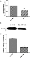
Fig 4. Regulation of S. xylosus IGPD by ISL.
(A) Effect of 1/2MIC of ISL on mRNA expression of hisB gene in S. xylosus. Data are expressed as means ± SDs. Significantly different means are indicated with asterisks (*) compared to the untreated control group (*p < 0.05, **p < 0.01, and ***p < 0.001). (B) Effect of 1/2MIC of ISL on the expression of IGPD protein in S. xylosus. (C) Determination of IGPD activity. Wild-type strain grown in the presence of 1/2-MICs of ISL. S. xylosus ATCC700404 served as control. Data are expressed as means ± SDs. Significantly different means are indicated with asterisks (*) compared to the untreated control group (*p < 0.05, **p < 0.01, and ***p < 0.001).
Fig 5
Fig 5. Determination of S. xylosus histidine content.
(A) The HPLC chromatograms of bacterial extract and histidine standard solution. I) The HPLC chromatograms of bacterial extract (black, retention time 14.781 min) and histidine standard solution (red, retention time 14.753 min). III and V) S. xylosus (wild-type strain or mutant strain) grown in the presence of 1/2 MIC of ISL. II and IV) Untreated wild-type strain and mutant strain served as a control. (B) Peak area analysis of S. xylosus histidine content by HPLC. S. xylosus (wild-type strain or mutant strain) grown in the presence of 1/2 MIC of ISL. Untreated wild-type strain and mutant strain were used as control. Data are expressed as means ± SDs. Significant difference compared to the untreated control group are represented with asterisk (*) (*p < 0.05, **p < 0.01, and ***p < 0.001).
Fig 6
Fig 6. Kinetic analysis by BLI of the binding of ISL to IGPD.
Fig 7
Fig 7. Computer methodological analysis.
(A) Tertiary structure model of S. xylosus IGPD. (B) Determination of the 3D structure of the IGPD-ISL complex using a molecular modeling method. (C) The predicted binding mode of IGPD with the ISL is shown, with labeling of key residues in the binding sites.
Fig 8
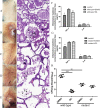
Fig 8. ISL provides protection against S. xylosus mastitis in a mouse model.
Lactating BALB/c mice were infected by injecting the canal glands with 1 × 10⁹ CFU bacterial cells. (A-E) The gross pathological changes (I) and histopathological analysis (II) of the mammary gland tissues at 48 h post-infection (n = 5) (Magnification: 600×). The infection group showed a large number of inflammatory factors and breast tissue destruction (arrowheads). Pathological abnormalities were significantly reduced by 250 μg/kg ISL in infected mice compared with DMSO treated mice. (F, G) ISL reduces the inflammatory response in infected mice. The levels of cytokines, including IL-6, and TNF-α, in the mammary gland tissues of infected mice were evaluated by ELISA (n = 5) (*p < 0.05, **p < 0.01, and ***p < 0.001). (H) The effect of the ISL on the bacterial burden in infected mice. Mammary gland tissues were collected, homogenized, and plated on TSB agar plates for the assessment of the bacterial burden (n = 5) (*p < 0.05, **p < 0.01, and ***p < 0.001). Results are reported as mean ± SD.

References

    1. Mushtaq S, Shah AM, Shah A, Lone SA, Hussain A, Hassan QP, et al. Bovine mastitis: An appraisal of its alternative herbal cure. Microbial Pathogenesis. 2018;114:357–61. 10.1016/j.micpath.2017.12.024 WOS:000426226700057. - DOI - PubMed
    1. Pyorala S, Taponen S. Coagulase-negative staphylococci-Emerging mastitis pathogens. Vet Microbiol. 2009;134(1–2):3–8. 10.1016/j.vetmic.2008.09.015 WOS:000263421900002. - DOI - PubMed
    1. Klimiene I, Virgailis M, Pavilonis A, Siugzdiniene R, Mockeliunas R, Ruzauskas M. Phenotypical and genotypical antimicrobial resistance of coagulase-negative staphylococci isolated from cow mastitis. Polish Journal of Veterinary Sciences. 2016;19(3):639–46. 10.1515/pjvs-2016-0080 WOS:000383528400024. - DOI - PubMed
    1. Akhaddar A, Elouennass M, Naama O, Boucetta M. Staphylococcus xylosus Isolated from an Otogenic Brain Abscess in an Adolescent. Surgical Infections. 2010;11(6):559–61. 10.1089/sur.2010.010 WOS:000285863400012. - DOI - PubMed
    1. Bochniarz M, Zdzisinska B, Wawron W, Szczubial M, Dabrowski R. Milk and serum IL-4, IL-6, IL-10, and amyloid A concentrations in cows with subclinical mastitis caused by coagulase-negative staphylococci. Journal of Dairy Science. 2017;100(12):9674–80. 10.3168/jds.2017-13552 WOS:000415926900017. - DOI - PubMed

Publication types

MeSH terms

Supplementary concepts