. 2019 Dec 20;15(12):e1008261.
doi: 10.1371/journal.pgen.1008261. eCollection 2019 Dec.

Restricted and non-essential redundancy of RNAi and piRNA pathways in mouse oocytes

Affiliations

Restricted and non-essential redundancy of RNAi and piRNA pathways in mouse oocytes

Eliska Taborska et al. PLoS Genet. .

Abstract

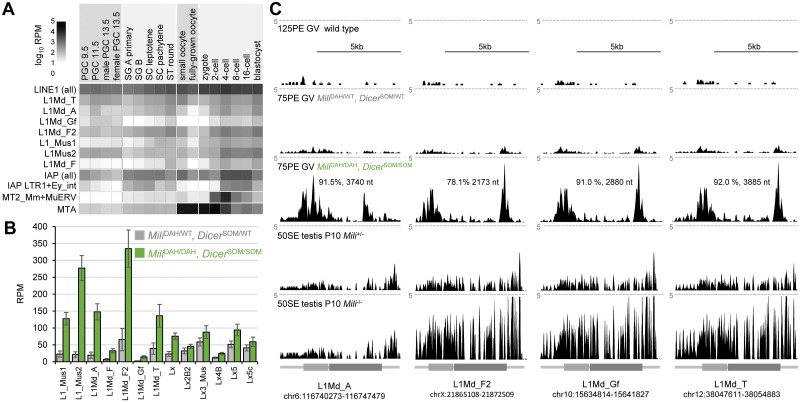
Germline genome defense evolves to recognize and suppress retrotransposons. One of defensive mechanisms is the PIWI-associated RNA (piRNA) pathway, which employs small RNAs for sequence-specific repression. The loss of the piRNA pathway in mice causes male sterility while females remain fertile. Unlike spermatogenic cells, mouse oocytes posses also RNA interference (RNAi), another small RNA pathway capable of retrotransposon suppression. To examine whether RNAi compensates the loss of the piRNA pathway, we produced a new RNAi pathway mutant DicerSOM and crossed it with a catalytically-dead mutant of Mili, an essential piRNA gene. Normal follicular and oocyte development in double mutants showed that RNAi does not suppress a strong ovarian piRNA knock-out phenotype. However, we observed redundant and non-redundant targeting of specific retrotransposon families illustrating stochasticity of recognition and targeting of invading retrotransposons. Intracisternal A Particle retrotransposon was mainly targeted by the piRNA pathway, MaLR and RLTR10 retrotransposons were targeted mainly by RNAi. Double mutants showed accumulations of LINE-1 retrotransposon transcripts. However, we did not find strong evidence for transcriptional activation and mobilization of retrotransposition competent LINE-1 elements suggesting that while both defense pathways are simultaneously expendable for ovarian oocyte development, yet another transcriptional silencing mechanism prevents mobilization of LINE-1 elements.


Conflict of interest statement

The authors have declared that no competing interests exist.

Figures

Fig 1
Fig 1. Retrotransposon-derived small RNAs.
Different retrotransposons are associated with different length distribution of small RNAs from mouse oocytes suggesting partially redundant repression of retrotransposons by endogenous RNAi and piRNA pathways. Based on RNA-seq data from Yang et al., 2016 [43].
Fig 2
Fig 2. DicerSOM mouse model.
(A) Schematic depiction of the 5’ gene structure of Dicer and DiceryΔMT and DicerSOM models. DicerSOM model was produced by removing ~6.5 kb of genomic DNA using CRISPR nucleases followed by homologous recombination with a construct encoding an HA-tag and exons 2–7. The resulting allele lacks introns 2 to 6 while encoding HA-tagged full-length Dicer. (B) DicerSOM mice express HA-tagged full-length Dicer. 40 μg of total protein lysate were loaded per lane and loading consistency was confirmed by Ponceau S staining of the Western blot membrane (lower panel). (C) DicerSOM protein has cytoplasmic expression. Shown are sections of seminiferous tubules stained with anti-HA antibody revealing cytoplasmic DicerSOM signal (red color) in spermatogonia of DicerSOM/SOM mice. DNA was stained with DAPI (blue color). (D) DicerSOM/SOM animals are born in a Mendelian ratio upon crossing DicerSOM/wt parents. (E) Analysis of Dicer expression by qPCR in oocytes of homozygous DicerΔMT and DicerSOM mutants. Bar colors indicate cDNA region amplified by primers localized in exons as described in the x-axis legend. DicerΔMT/ΔMT mice show loss of expression of the MTC-driven Dicer variant and a relative increase of expression from the downstream MTA LTR insertion. DicerSOM/SOM mice show complete loss of short Dicer isoform. Data from three biological replicates were normalized to oocytes from heterozygous littermates. Error bar = SD. (F) Analysis of Dicer expression in oocytes of wild-type animals and homozygous DicerΔMT and DicerSOM mutants by RNA-seq (three biological replicates). Depicted are RPKM values per exon. Error bar = SD.
Fig 3
Fig 3. DicerSOM/SOM animals phenocopy oocyte-specific Dicer knock-out.
(A) Oocytes of DicerSOM/SOM mice exhibit spindle defects like Dicer−/− or DicerΔMT/ΔMT oocytes [38, 39, 44]. Size bar = 10 μm. (B) Frequency of meiotic spindle defects in oocytes from DicerSOM/wt and DicerSOM/SOM mice. Numbers correspond to examined oocytes with each genotype. (C) mRNA expression of selected RNAi targets analyzed by qPCR performed as three biological replicates. B2m is a non-targeted gene. All mRNA levels are shown relative to Hprt1. (D) MA plots depicting transcriptome changes in DicerΔMT/ΔMT and DicerSOM/SOM oocytes relative to wild-type oocytes. (E) PCA analysis of RPKM transcriptome changes in oocytes of different mutants, which includes previously published data from Dicer and Ago mutants [31]. (F) Comparison of mRNA expression changes in DicerΔMT/ΔMT and DicerSOM/SOM oocytes (x-axis) with mRNA changes in oocytes with conditional Dicer knock-out (y-axis). (G) Correlation matrices of retrotransposon-derived transcriptome in different mutant oocytes. Elliptic shapes reflect sizes of inscribed correlation coefficients for easier visual navigation.
Fig 4
Fig 4. RNAi (DicerSOM/SOM) and piRNA (MiliDAH/DAH) knock-out analysis.
(A) Representative histological sections of ovaries stained with hematoxylin and eosin. Abbreviations: CL–corpus luteum, EA—early antral follicle, AF—antral follicle. (B) Ovarian weight. Ten ovaries from five 14–19 weeks-old females were analyzed per genotype. (C) Yield of fully-grown germinal vesicle (GV)-intact oocytes per animal. Seven 10–16 weeks old females per genotype were analyzed. (D) Analysis of follicles in ovaries of mutant animals. Follicular content has been normalized per average ovarian section area per animal. Six ovaries from three animals were analyzed. Error bar = SD.
Fig 5
Fig 5. Transcriptional changes in oocytes lacking RNAi and piRNA pathways.
(A) qPCR analysis of selected retrotransposons in mutant oocytes. Retrotransposon RNA levels are shown relative to that in heterozygous (MiliDAH/WT, DicerSOM/WT) oocytes. Three biological replicates were perfomed. Error bar = SD. (B) PCA analysis of RPKM transcriptome changes in oocytes with mutated RNAi (MiliDAH/WT, DicerSOM/SOM), piRNA (MiliDAH/DAH, DicerSOM/WT) or both pathways (MiliDAH/DAH, DicerSOM/SOM). Oocytes from heterozygous littermates (MiliDAH/WT, DicerSOM/WT) served as controls. (C) MA plot depicting transcriptome changes in double mutant oocytes relative to RNAi pathway mutant oocytes. MA plots depicting transcriptome changes of pathway mutants relative to heterozygous control are provided in S3 Fig. Transcripts with significantly higher and lower transcript levels are shown in red and blue, respectively and are provided in the Supplementary file 1. Transcripts from a tandem duplication cluster at chromosome 12 are depicted as triangles. One such a transcript was chosen for display in the panel E. Transcript labeled with D is associated with a Mili-dependent small RNA cluster. (D) A UCSC browser snapshot of a region producing Mili-dependent and Mili-independent small RNAs. The locus encodes non-coding RNAs (Gm476432 and Gm17821), which show increased abundance in double mutants. The upper part shows RNA-seq data from oocytes with the four genotypes. Dashed horizontal lines depict expression level corresponding to 100 counts per million (CPM). The lower two samples show small RNAs from wild-type and Mili-mutant postnatal ovaries (day 20) from Kabayama et al. [21]. The MiliDAH/DAH mutant is the same one as in our study. The bottom part depicts distribution of repetitive elements along the locus with a blue arrow pointing to the LTRIS_Mm inverted repeat giving rise to Mili-independent 21–23 nt small RNAs. (E) An example of one of the paralogs from the tandem duplication cluster at chromosome 12, which exhibit increased transcript levels in double mutants. The upper part shows the tandem duplication cluster at chromosome 12, the lower half shows a snapshot from a UCSC browser with scaled RNA-seq data from different mutants. Expression analysis data from UCSC browser are organized as in the panel D. The bottom part depicts distribution of SINE, LINE and LTR retrotransposons and MurSatRep1 repeat along the locus. (F) RNA abundance of selected retrotransposon types from Repeatmasker annotation in mutant oocytes are displayed relative to heterozygous (MiliDAH/WT, DicerSOM/WT) controls.
Fig 6
Fig 6. Retrotransposon expression in the female germline.
(A) Relative retrotransposon expression during the germline cycle. The heatmap was produced as described previously [46] using published expression data [, –50]. Depicted L1 and IAP retrotransposon subfamilies have full-length intact retrotransposon insertions in C57Bl/6 genome. (B) Selected L1 retrotransposon contribution to transcriptomes of double mutants (MiliDAH/DAH, DicerSOM/SOM) and controls (MiliDAH/WT, DicerSOM/WT). Four biological replicates. Error bar = SD. (C) Most expressed intact L1 retrotransposons from the four most abundant subfamilies. Shown are UCSC genome browser snapshots of specific L1 insertions with the highest coverage by RNA-seq reads in double mutants. The samples (from the top) depict merged data from 125 nucleotide paired-end (125PE) sequencing of normal full-grown GV oocytes from Horvat et al. [54], 75 nucleotide paired-end (75PE) sequencing of controls (MiliDAH/WT, DicerSOM/WT) and double mutants (MiliDAH/DAH, DicerSOM/SOM) from this study, and 50 nucleotide single-end (50SE) sequencing of total transcriptome of 10 days old wild-type and Mili-–/-– testes [28]. All RNA-seq data were scaled and are shown at the same scale 5 CPM indicated by the dashed line. L1 schemes are depicted with ORF1 and ORF2 as lighter and darker grey rectangle, respectively.

References

    1. Bourque G, Burns KH, Gehring M, Gorbunova V, Seluanov A, Hammell M, et al. Ten things you should know about transposable elements. Genome Biol. 2018;19(1):199 10.1186/s13059-018-1577-z . - DOI - PMC - PubMed
    1. Craig NL, Chandler M, Gellert M, Lambowitz AM, Rice PA, Sandmeyer SB. Mobile DNA III. Craig NL, editor: AMS press; 2015.
    1. International Human Genome Sequencing Consortium, Lander ES, Linton LM, Birren B, Nusbaum C, Zody MC, et al. Initial sequencing and analysis of the human genome. Nature. 2001;409(6822):860–921. 10.1038/35057062 . - DOI - PubMed
    1. Mouse Genome Sequencing Consortium, Waterston RH, Lindblad-Toh K, Birney E, Rogers J, Abril JF, et al. Initial sequencing and comparative analysis of the mouse genome. Nature. 2002;420(6915):520–62. 10.1038/nature01262 . - DOI - PubMed
    1. Kazazian HH Jr. Mobile elements and disease. Curr Opin Genet Dev. 1998;8(3):343–50. 10.1016/s0959-437x(98)80092-0 . - DOI - PubMed

Publication types