

Multiple calcaneal fibrous dysplasia: A case report
- PMID: 31861003
- PMCID: PMC6940136
- DOI: 10.1097/MD.0000000000018389
Multiple calcaneal fibrous dysplasia: A case report
Abstract
Rationale: Fibrous dysplasia (FD) is a benign bone tumor due to developmental failure in the process of primitive bone remodeling to mature lamellar bone. The most common locations of monostotic FD of the extremity bones are the proximal femur, tibia, humerus and the radius. FD in the calcaneus is extremely rare and usually manifests clinically as a single bone lesion. Moreover, no research has reported on multiple lesions in calcaneal FD.
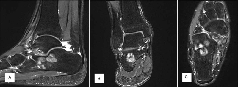
Patient concerns: We report a 21-year-old man presented to our institution with pain upon walking for 2 months.
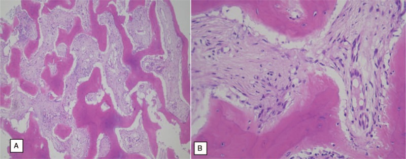
Diagnoses: We diagnosed the patient with multiple calcaneal FD through histologic examination of the excised biopsy that revealed cellular, spindly stroma and woven bone without osteoblastic rimming resembling Chinese characters INTERVENTIONS:: Plain X-ray, computed tomography, magnetic resonance imaging and histologic examination. An excisional biopsy with extended curettage and bone grafting with allogenous bone and autogenous bone marrow aspirate concentrate were performed.
Outcomes: No complications developed after surgery and during serial follow-ups at 3, 6 and 12 months. At a postoperative 12-month follow-up, a plain radiogram showed a well-consolidated bone graft in the lesions.
Lessons: Calcaneal FD is rare disease entity. This case can help guide clinical decision-making in the future.
Conflict of interest statement
The authors have no conflicts of interest to disclose.
Figures


References
-
- Bartley J, Munroe SM, Ward RA. Fibrous dysplasia in the calcaneus. Foot Ankle Spec 2017;10:72–4. - PubMed
-
- Isefuku S, Hatori M, Ehara S, et al. Fibrous dysplasia arising from the calcaneus. Tohoku J Exp Med 1999;189:227–32. - PubMed
-
- Pandy S. Fibrous dysplasia of the calcaneum. Report of three cases. Int Surg 1971;55:119–22. - PubMed
-
- Parekh SG, Donthineni-rao R, Ricchetti E, et al. Fibrous dysplasia. J Am Acad Orthop Surg 2004;12:305–13. - PubMed
Publication types
MeSH terms
LinkOut - more resources
Full Text Sources
