

Pericytes in Microvessels: From "Mural" Function to Brain and Retina Regeneration
- PMID: 31861092
- PMCID: PMC6940987
- DOI: 10.3390/ijms20246351
Pericytes in Microvessels: From "Mural" Function to Brain and Retina Regeneration
Abstract
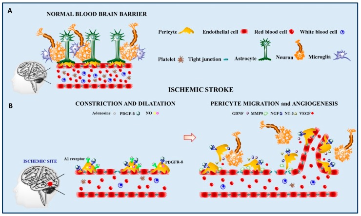
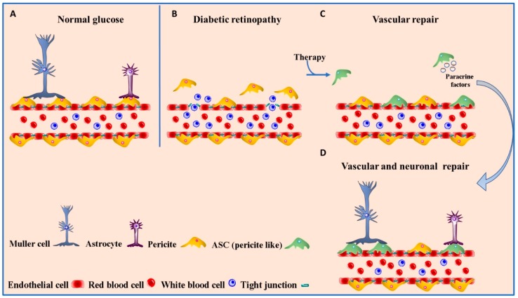
Pericytes are branched cells located in the wall of capillary blood vessels that are found throughout the body, embedded within the microvascular basement membrane and wrapping endothelial cells, with which they establish a strong physical contact. Pericytes regulate angiogenesis, vessel stabilization, and contribute to the formation of both the blood-brain and blood-retina barriers by Angiopoietin-1/Tie-2, platelet derived growth factor (PDGF) and transforming growth factor (TGF) signaling pathways, regulating pericyte-endothelial cell communication. Human pericytes that have been cultured for a long period give rise to multilineage progenitor cells and exhibit mesenchymal stem cell (MSC) features. We focused our attention on the roles of pericytes in brain and ocular diseases. In particular, pericyte involvement in brain ischemia, brain tumors, diabetic retinopathy, and uveal melanoma is described. Several molecules, such as adenosine and nitric oxide, are responsible for pericyte shrinkage during ischemia-reperfusion. Anti-inflammatory molecules, such as IL-10, TGFβ, and MHC-II, which are increased in glioblastoma-activated pericytes, are responsible for tumor growth. As regards the eye, pericytes play a role not only in ocular vessel stabilization, but also as a stem cell niche that contributes to regenerative processes in diabetic retinopathy. Moreover, pericytes participate in melanoma cell extravasation and the genetic ablation of the PDGF receptor reduces the number of pericytes and aberrant tumor microvessel formation with important implications for therapy efficacy. Thanks to their MSC features, pericytes could be considered excellent candidates to promote nervous tissue repair and for regenerative medicine.
Keywords: MSC features; Pericytes; brain and retina repair; brain tumor; diabetic retinopathy; microvessels; ocular diseases; stroke.
Conflict of interest statement
The authors declare no conflict of interest.
Figures
References
-
- Diaz-Flores L., Gutierrez R., Madrid J.F., Varela H., Valladares F., Acosta E., Martin-Vasallo P., Diaz-Flores L. Pericytes. Morphofunction, interactions and pathology in a quiescent and activated mesenchymal cell niche. Histol Histopathol. 2009;24:909–969. - PubMed
-
- Zimmermann K.W. Der feinere Bau der Blutcapillaren. Z. Anat. Entwickl. 1923;68:29–109. doi: 10.1007/BF02593544. - DOI
Publication types
MeSH terms
LinkOut - more resources
Full Text Sources
Research Materials
Miscellaneous
