

Site-Specific 111In-Radiolabeling of Dual-PEGylated Porous Silicon Nanoparticles and Their In Vivo Evaluation in Murine 4T1 Breast Cancer Model
- PMID: 31861119
- PMCID: PMC6969933
- DOI: 10.3390/pharmaceutics11120686
Site-Specific 111In-Radiolabeling of Dual-PEGylated Porous Silicon Nanoparticles and Their In Vivo Evaluation in Murine 4T1 Breast Cancer Model
Abstract
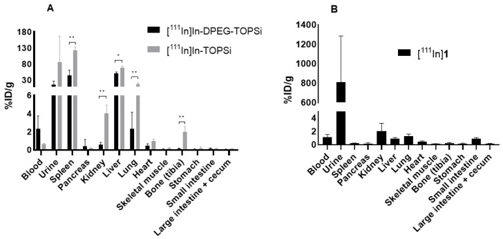
Polyethylene glycol (PEG) has been successfully used for improving circulation time of several nanomaterials but prolonging the circulation of porous silicon nanoparticles (PSi NPs) has remained challenging. Here, we report a site specific radiolabeling of dual-PEGylated thermally oxidized porous silicon (DPEG-TOPSi) NPs and investigation of influence of the PEGylation on blood circulation time of TOPSi NPs. Trans-cyclooctene conjugated DPEG-TOPSi NPs were radiolabeled through a click reaction with [111In]In-DOTA-PEG4-tetrazine (DOTA = 1,4,7,10-tetraazacyclododecane-1,4,7,10-tetraacetic acid) and the particle behavior was evaluated in vivo in Balb/c mice bearing 4T1 murine breast cancer allografts. The dual-PEGylation significantly prolonged circulation of [111In]In-DPEG-TOPSi particles when compared to non-PEGylated control particles, yielding 10.8 ± 1.7% of the injected activity/g in blood at 15 min for [111In]In-DPEG-TOPSi NPs. The improved circulation time will be beneficial for the accumulation of targeted DPEG-TOPSi to tumors.
Keywords: IEDDA; SPECT; click chemistry; indium-111; porous silicon.
Conflict of interest statement
The authors declare no conflict of interest.
Figures


Similar articles
-
Impact of Pore Size and Surface Chemistry of Porous Silicon Particles and Structure of Phospholipids on Their Interactions.ACS Biomater Sci Eng. 2018 Jul 9;4(7):2308-2313. doi: 10.1021/acsbiomaterials.8b00343. Epub 2018 Jun 14. ACS Biomater Sci Eng. 2018. PMID: 30159385 Free PMC article.
-
Tailored Dual PEGylation of Inorganic Porous Nanocarriers for Extremely Long Blood Circulation in Vivo.ACS Appl Mater Interfaces. 2016 Dec 7;8(48):32723-32731. doi: 10.1021/acsami.6b12481. Epub 2016 Nov 23. ACS Appl Mater Interfaces. 2016. PMID: 27934159
-
Effect of surface chemistry of porous silicon microparticles on glucagon-like peptide-1 (GLP-1) loading, release and biological activity.Int J Pharm. 2013 Sep 15;454(1):67-73. doi: 10.1016/j.ijpharm.2013.06.063. Epub 2013 Jul 5. Int J Pharm. 2013. PMID: 23834832
-
Improved stability and biocompatibility of nanostructured silicon drug carrier for intravenous administration.Acta Biomater. 2015 Feb;13:207-15. doi: 10.1016/j.actbio.2014.11.019. Epub 2014 Nov 20. Acta Biomater. 2015. PMID: 25463492
-
Impact of PEGylated Nanoparticles on Tumor Targeted Drug Delivery.Curr Pharm Des. 2018;24(28):3283-3296. doi: 10.2174/1381612824666180730161721. Curr Pharm Des. 2018. PMID: 30062957 Review.
Cited by
-
Radiolabeled nanomaterial for cancer diagnostics and therapeutics: principles and concepts.Cancer Nanotechnol. 2023;14(1):15. doi: 10.1186/s12645-023-00165-y. Epub 2023 Feb 27. Cancer Nanotechnol. 2023. PMID: 36865684 Free PMC article. Review.
-
Recent Developments in Porous Silicon Nanovectors with Various Imaging Modalities in the Framework of Theranostics.ChemMedChem. 2022 May 18;17(10):e202200004. doi: 10.1002/cmdc.202200004. Epub 2022 Mar 18. ChemMedChem. 2022. PMID: 35212460 Free PMC article. Review.
-
Methods for Radiolabelling Nanoparticles: SPECT Use (Part 1).Biomolecules. 2022 Oct 20;12(10):1522. doi: 10.3390/biom12101522. Biomolecules. 2022. PMID: 36291729 Free PMC article. Review.
-
Click Chemistry and Radiochemistry: An Update.Bioconjug Chem. 2023 Nov 15;34(11):1925-1950. doi: 10.1021/acs.bioconjchem.3c00286. Epub 2023 Sep 22. Bioconjug Chem. 2023. PMID: 37737084 Free PMC article. Review.
-
In Vivo Imaging of [60]Fullerene-Based Molecular Spherical Nucleic Acids by Positron Emission Tomography.Mol Pharm. 2023 Oct 2;20(10):5043-5051. doi: 10.1021/acs.molpharmaceut.3c00370. Epub 2023 Aug 2. Mol Pharm. 2023. PMID: 37531591 Free PMC article.
References
-
- Dogra P., Adolphi N.L., Wang Z., Lin Y.-S., Butler K.S., Durfee P.N., Croissant J.G., Noureddine A., Coker E.N., Bearer E.L., et al. Establishing the effects of mesoporous silica nanoparticle properties on in vivo disposition using imaging-based pharmacokinetics. Nat. Commun. 2018;9:4551. doi: 10.1038/s41467-018-06730-z. - DOI - PMC - PubMed
Grants and funding
LinkOut - more resources
Full Text Sources
