

Imaging Modalities for Diagnosis of Deep Pelvic Endometriosis: Comparison between Trans-Vaginal Sonography, Rectal Endoscopy Sonography and Magnetic Resonance Imaging. A Head-to-Head Meta-Analysis
- PMID: 31861142
- PMCID: PMC6963762
- DOI: 10.3390/diagnostics9040225
Imaging Modalities for Diagnosis of Deep Pelvic Endometriosis: Comparison between Trans-Vaginal Sonography, Rectal Endoscopy Sonography and Magnetic Resonance Imaging. A Head-to-Head Meta-Analysis
Abstract
Objectives: A meta-analysis, with a head-to-head approach, was carried out to compare the three most common techniques for a deep pelvic endometriosis (DPE) diagnosis. We focused on: transvaginal-sonography (TVS), magnetic-resonance imaging (MRI), and rectal-endoscopy-sonography (RES).
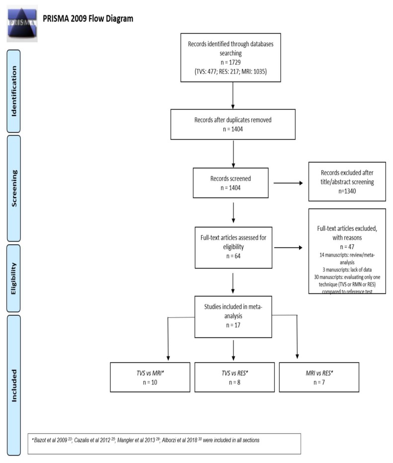
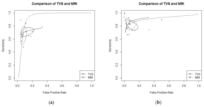
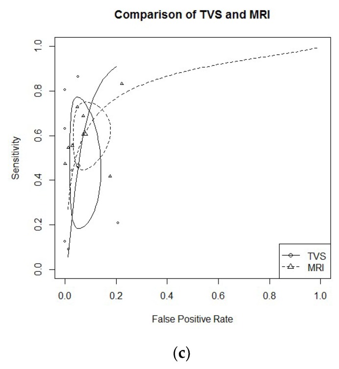
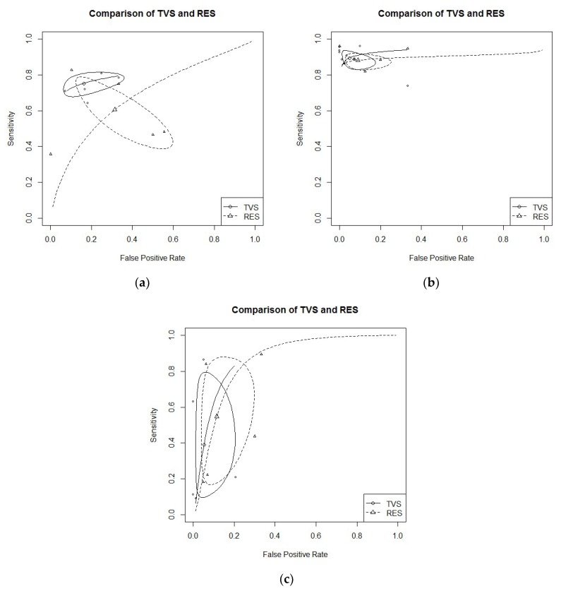
Methods: Electronic databases were searched from their inception until December 2018. All prospective and well-defined retrospective studies carried out in tertiary referral centers were considered. This review was reported following the Preferred Reporting Items for Systematic Reviews and Meta-Analyses (PRISMA) and Synthesizing Evidence from Diagnostic Accuracy Tests (SEDATE) guidelines. We considered only papers in which at least two imaging modalities were compared in the same set of patients (head-to-head approach). Meta-analysis of diagnostic test accuracy (DTA) was performed separately for each location of interest. Bivariate or univariate approach has been applied when appropriate. We analyze the DTA of TVS vs. MRI, TVS vs. RES, and MRI vs. RES.
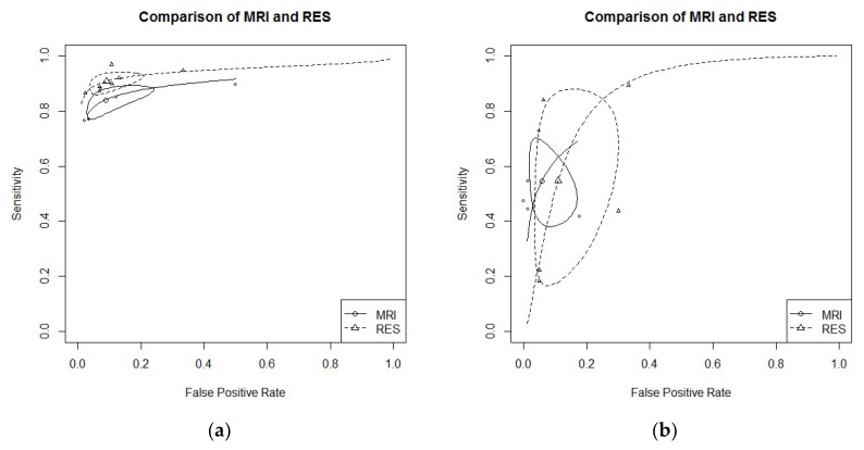
Results: Our meta-analysis (17 studies included) showed high-to-moderate DTA of TVS for all endometriosis locations (apart from recto-vaginal septum (RVS)) that were not statistically different from MRI and RES for those localized in the posterior compartment. RES results were more accurate than MRI for RS lesions but less accurate than TVS for other pelvic locations, except for RVS.
Conclusions: All approaches provide good accuracy with specific strong points. Ultrasonography demonstrated a diagnostic accuracy not inferior to MRI and RES; therefore, it must be considered the primary approach for DPE diagnosis. MRI has to be considered as a valuable approach in settings where highly skilled sonographers are not available. Keypoints: (1) We confirmed the non-inferiority of TVS compared to MRI and RES for the diagnosis of specific pelvic anatomic location of endometriosis lesions. (2) Ultrasonography could be considered the primary approach for DPE diagnosis (less invasive than RES and less expensive than MRI). (3) MRI has to be considered as a valuable approach in settings where skilled sonographers are not available.
Keywords: diagnostic tests; early diagnosis; endometriosis; magnetic resonance imaging; pelvis; sensitivity and specificity; ultrasonography.
Conflict of interest statement
The authors declare no conflict of interest.
Figures
References
-
- Noventa M., Saccardi C., Litta P., Vitagliano A., D′Antona D., Abdulrahim B., Duncan A., Alexander-Sefre F., Aldrich C.J., Quaranta M., et al. Ultrasound techniques in the diagnosis of deep pelvic endometriosis: Algorithm based on a systematic review and meta-analysis. Fertil. Steril. 2015;104:366–383. doi: 10.1016/j.fertnstert.2015.05.002. - DOI - PubMed
-
- Nnoaha K.E., Hummelshoj L., Webster P., d’Hooghe T., de Cicco Nardone F., de Cicco Nardone C., Jenkinson C., Kennedy S.H., Zondervan K.T. World Endometriosis Research Foundation Global Study of Women’s Health consortium. Reprint of: Impact of endometriosis on quality of life and work productivity: A multicenter study across ten countries. Fertil. Steril. 2019;112:e137–e152. doi: 10.1016/j.fertnstert.2019.08.082. - DOI - PubMed
-
- Scioscia M., Bruni F., Ceccaroni M., Steinkasserer M., Stepniewska Minelli L. Distribution of endometriotic lesions in endometriosis stage IV supports the menstrual reflux theory and requires specific preoperative assessment and therapy. Acta Obstet. Gynecol. Scand. 2011;90:136–139. doi: 10.1111/j.1600-0412.2010.01008.x. - DOI - PubMed
Publication types
LinkOut - more resources
Full Text Sources
