. 2019 Dec 17;20(24):6363.
doi: 10.3390/ijms20246363.

Cardiac Extracellular Vesicles (EVs) Released in the Presence or Absence of Inflammatory Cues Support Angiogenesis in Different Manners

Affiliations

Cardiac Extracellular Vesicles (EVs) Released in the Presence or Absence of Inflammatory Cues Support Angiogenesis in Different Manners

Christien Madlen Beez et al. Int J Mol Sci. .

Abstract

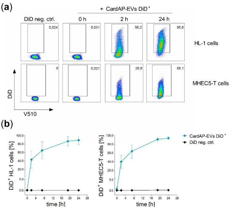
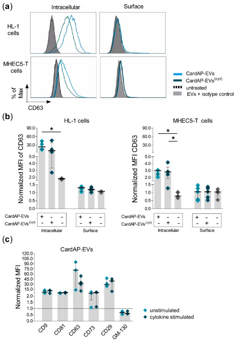
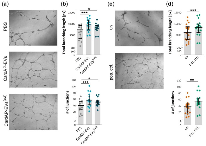
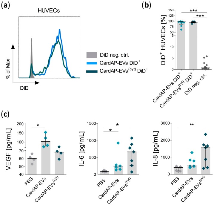
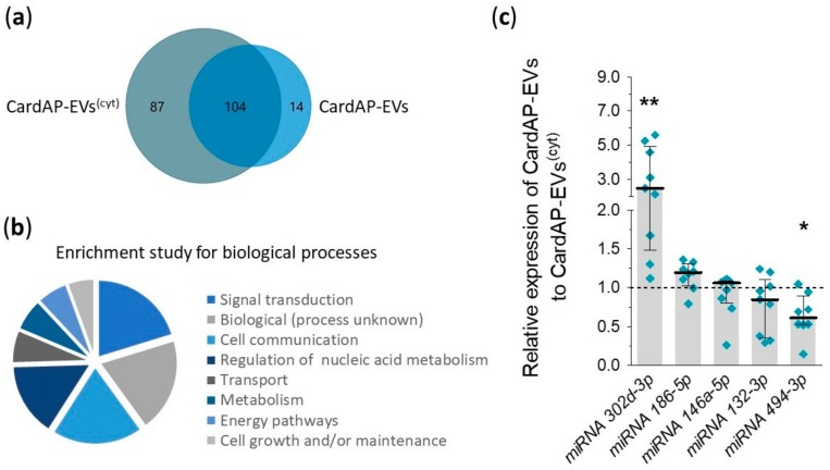
Cells release extracellular vesicles (EVs) to communicate in a paracrine manner with other cells, and thereby influence processes, such as angiogenesis. The conditioned medium of human cardiac-derived adherent proliferating (CardAP) cells was recently shown to enhance angiogenesis. To elucidate whether their released EVs are involved, we isolated them by differential centrifugation from the conditioned medium derived either in the presence or absence of a pro-inflammatory cytokine cocktail. Murine recipient cells internalized CardAP-EVs as determined by an intracellular detection of human proteins, such as CD63, by a novel flow cytometry method for studying EV-cell interaction. Moreover, endothelial cells treated for 24 h with either unstimulated or cytokine stimulated CardAP-EVs exhibited a higher tube formation capability on Matrigel. Interestingly, unstimulated CardAP-EVs caused endothelial cells to release significantly more vascular endothelial growth factor and interleukin (IL)-6, while cytokine stimulated CardAP-EVs significantly enhanced the release of IL-6 and IL-8. By nCounter® miRNA expression assay (NanoString Technologies) we identified microRNA 302d-3p to be enhanced in unstimulated CardAP-EVs compared to their cytokine stimulated counterparts, which was verified by quantitative polymerase chain reaction. This study demonstrates that both CardAP-EVs are pro-angiogenic by inducing different factors from endothelial cells. This would allow to select potent targets for a safe and efficient therapeutic application.

Keywords: VEGF; angiogenesis; cardiac therapy; extracellular vesicles; intracellular uptake; regeneration.


Conflict of interest statement

M. Sittinger is a shareholder of CellServe GmbH (Berlin, Germany) and BioRetis GmbH (Berlin, Germany). CellServe owns a license for CardAP cells. M. Haag and M. Sittinger are investors of patent EP000002129774A1 “Cells for heart treatment”. The remaining authors report no competing financial interests.

Figures

Figure 1
Figure 1
CardAP-EVs (EVs from cardiac derived adherent proliferating cells) interact equally with cardiomyocytes and cardiac endothelial cells. DiD labelled CardAP-EVs (CardAP-EVs DiD+) as well as a DiD negative control (DiD neg. ctrl.) were applied to cultured HL-1 cells or MHEC5-T cells (2 × 10⁵ cells/well). At different time points (0, 2, 7, 19, and 24 h) cells were washed twice with PBS, harvested and labelled with a dead/viable marker (V510) for 20 min and fixed with 0.5% PFA after a washing step. All samples were analyzed by flow cytometry at the Canto II. (a): Representative dot plots are shown for different time points of HL-1 cells (upper raw) as well as for MHEC5-T cells (lower raw) treated with CardAP-EVs or treated for 24 h with the control. (b): The frequency of DiD+ HL-1 cells (right, n = 4, 4 different CardAP donors) and DiD+ MHEC5-T cells (left, n = 4–6, 3 different CardAP donors) is shown in relation to the time of incubation (h) as median with interquartile range for treatment with either CardAP-EVs (blue) or control (black).
Figure 2
Figure 2
CardAP-EVs are internalized by cardiac murine cells. Human CardAP-EVs either unstimulated (CardAP-EVs) or cytokine stimulated (CardAP-EVs(cyt)) were applied to cultured murine HL-1 cells or MHEC5-T cells (2 × 10⁵ cells/well). Additionally, an untreated murine cell control was included. After 24 h, these cells were harvested and stained with anti-human fluorescently labelled antibodies for CD63, using either an intracellular or a cell surface staining protocol. Stained samples were measured by flow cytometry at the Canto II. (a): Representative histograms are shown for HL-1 cells (upper row) and MHEC5-T cells (lower row) for human CD63 via intracellular (left) or surface (right) staining in comparison to untreated and isotype control (EVs + isotype ctrl.) (b): The mean fluorescent intensity (MFI) for human CD63 was normalized to an unstained control (intracellular or surface staining) and the normalized MFI is shown as median with interquartile range for both staining conditions as well as different treatments for HL-1 cells (left, n = 4, 3 different CardAP donors) and MHEC5-T cells (right, n = 5, 3 different CardAP donors). (c): The presence of human markers (CD9, CD81, CD63, CD73, CD29) and the Golgi matrix protein (GM-130) on CardAP-EVs was verified by flow cytometry of EVs bound to sulfate/aldehyde beads. The normalized MFI are shown for all markers as median with interquartile range (n = 3–4, 3 different CardAP donors). Statistical analysis was performed by Kruskal-Wallis test (with Dunn’s post hoc test; * p < 0.05).
Figure 3
Figure 3
Tube formation capabilities of HUVECs is enhanced by both unstimulated and cytokine stimulated CardAP-EVs. 24 h prior to an assay, HUVECs were treated with 6 µg/mL of either unstimulated (CardAP-EVs) or cytokine stimulated CardAP-EVs (CardAP-EVs(cyt)), PBS in corresponding volumes of CardAP-EVs, or left untreated (un). The next day, HUVECs were harvested and applied to a Matrigel-coated well for 20 h. As a positive control, HUVECs were treated in parallel with 10 ng/mL VEGF. The tube formation was documented by light microscopy and pictures were analyzed with the help of the ImageJ Angiogenesis Plugin. (a): Representative pictures are shown for HUVECs treated with PBS, unstimulated and cytokine stimulated CardAP-EVs with a scale bar representing 500 µm. (b): The quantitative analysis of the tube formation shows a significant increase of the total branching length (upper graph) and the number of junctions (lower graph) for unstimulated as well as cytokine stimulated CardAP-EVs. Individual data points are shown and summarized as median with interquartile range (n = 16, 6 different CardAP donors). (c): Representative pictures are shown for HUVECs without any treatment (un) or for HUVECs treated with 10 ng/mL VEGF (pos. ctrl.). The scale bar represents 500 µm. (d): The total branching length (upper graph) as well as the number of junctions (lower graph) is significantly increased by the application of VEGF versus the untreated samples. Individual data points are shown and summarized as median with interquartile range (n = 17). Statistical analysis for three groups was performed by repeated measures ANOVA (Bonferroni post hoc test, *** p < 0.001, * p < 0.05). Statistical analysis of two groups was performed by paired T test (*** p < 0.001, ** p < 0.01).
Figure 4
Figure 4
The interaction of unstimulated and cytokine stimulated CardAP-EVs with HUVECs triggers the release of different pro-angiogenic factors. Cultured HUVECs (1.9 × 10⁵ cells/well) were treated with DiD labelled CardAP-EVs or with a DiD negative control. After 24 h, HUVECs were washed twice with PBS, harvested, and labelled with a dead/viable marker (V510) for 20 min and fixed with 0.5% PFA after a washing step. All samples were analyzed by flow cytometry at the Canto II. (a): Representative histograms are shown for HUVECs treated with the DiD negative control (DiD neg. ctrl.), DiD labelled unstimulated CardAP-EVs (CardAP-EVs DiD+) or cytokine stimulated CardAP-EVs (CardAP-EVs(cyt) DiD+) (b): The frequency of DiD+ HUVECs (n = 6–13, 4 different CardAP donors) is shown as median with interquartile range for the three different treatments. (c): HUVECs were treated with 6 µg/mL of either unstimulated (EVs), cytokine stimulated CardAP-EVs (EVs(cyt)) or PBS in volumes corresponding to that of the CardAP-EVs. The next day, HUVECs were washed twice and fresh medium was applied for 24 h. Then, the medium was collected for detecting IL-6, IL-8, and VEGF by ELISA. The cell correction factor was determined by crystal violet staining. The cytokine concentrations in relation to the cell numbers are shown for VEGF (n = 4, 4 different CardAP donors), IL-6, and IL-8 (both n = 7, 4 different CardAP donors) as median with interquartile range. Statistical analysis was performed by Friedman test (with Dunn’s post hoc test; *** p < 0.001, ** p < 0.01, * p < 0.05).
Figure 5
Figure 5
Different composition of miRNAs between unstimulated and cytokine stimulated CardAP-EVs. RNA was isolated from unstimulated and cytokine stimulated CardAP-EVs and used for nCounter® miRNA expression assay as stated in the materials and methods section. (a,b): The derived data was normalized and used for further analysis by FunRich Analysis for a comparison of observed miRNAs between both conditions illustrated as Venn diagram (a) or as general analysis of observed miRNAs for their involvement in biological processes as displayed as pie chart for the top eight processes (b). (c): Five miRNAs (miR302d, miR186-5p, miR146a-5p, miR132-3p, and miR494-3p) identified in the global nCounter® miRNA expression analysis, were validated by qPCR. Values of unstimulated CardAP-EVs were normalized to the cytokine stimulated CardAP-EVs (set to 1, black dotted line) by means of ΔΔCt analysis. The relative expression is shown as median with interquartile range (n = 8–9, 3 different CardAP donors). Statistical analysis was performed with a Wilcoxon signed rank test (** p < 0.01, * p< 0.05).

Similar articles

Cited by

References

    1. Théry C., Ostrowski M., Segura E. Membrane vesicles as conveyors of immune responses. Nat. Rev. Immunol. 2009;9:581–593. doi: 10.1038/nri2567. - DOI - PubMed
    1. Yoon Y.J., Kim O.Y., Gho Y.S. Extracellular vesicles as emerging intercellular communicasomes. BMB Rep. 2014;47:531–539. doi: 10.5483/BMBRep.2014.47.10.164. - DOI - PMC - PubMed
    1. Alfaro M.P., Saraswati S., Young P.P. Molecular Mediators of Mesenchymal Stem Cell Biology. Vitam. Horm. 2011;87:39–59. - PMC - PubMed
    1. Collino F., Bruno S., Deregibus M.C., Tetta C., Camussi G. MicroRNAs and Mesenchymal Stem Cells. Vitam. Horm. 2011;87:291–320. - PubMed
    1. Loewenstein W.R. Junctional intercellular communication: the cell-to-cell membrane channel. Physiol. Rev. 1981;61:829–913. doi: 10.1152/physrev.1981.61.4.829. - DOI - PubMed

LinkOut - more resources