. 2019 Dec 18;9(1):1.
doi: 10.3390/antiox9010001.

Peroxiredoxin II Maintains the Mitochondrial Membrane Potential against Alcohol-Induced Apoptosis in HT22 Cells

Affiliations

Peroxiredoxin II Maintains the Mitochondrial Membrane Potential against Alcohol-Induced Apoptosis in HT22 Cells

Mei-Hua Jin et al. Antioxidants (Basel). .

Abstract

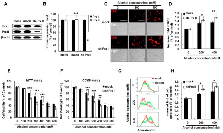
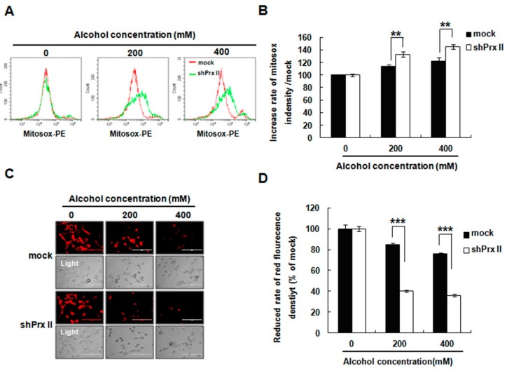
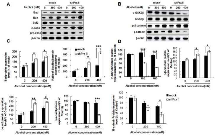
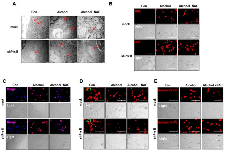
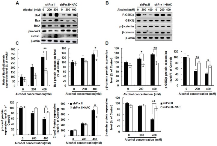
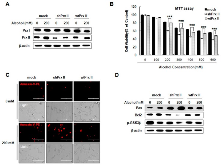
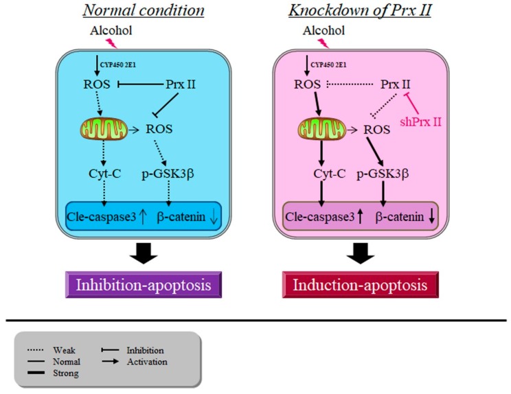
Excessive alcohol intake can significantly reduce cognitive function and cause irreversible learning and memory disorders. The brain is particularly vulnerable to alcohol-induced ROS damage; the hippocampus is one of the most sensitive areas of the brain for alcohol neurotoxicity. In the present study, we observed significant increasing of intracellular ROS accumulations in Peroxiredoxin II (Prx II) knockdown HT22 cells, which were induced by alcohol treatments. We also found that the level of ROS in mitochondrial was also increased, resulting in a decrease in the mitochondrial membrane potential. The phosphorylation of GSK3β (Ser9) and anti-apoptotic protein Bcl2 expression levels were significantly downregulated in Prx II knockdown HT22 cells, which suggests that Prx II knockdown HT22 cells were more susceptible to alcohol-induced apoptosis. Scavenging the alcohol-induced ROS with NAC significantly decreased the intracellular ROS levels, as well as the phosphorylation level of GSK3β in Prx II knockdown HT22 cells. Moreover, NAC treatment also dramatically restored the mitochondrial membrane potential and the cellular apoptosis in Prx II knockdown HT22 cells. Our findings suggest that Prx II plays a crucial role in alcohol-induced neuronal cell apoptosis by regulating the cellular ROS levels, especially through regulating the ROS-dependent mitochondrial membrane potential. Consequently, Prx II may be a therapeutic target molecule for alcohol-induced neuronal cell death, which is closely related to ROS-dependent mitochondria dysfunction.

Keywords: Prx II; ROS; alcohol; neuronal cell; oxidative damage.


Conflict of interest statement

The authors declare that there are no conflicts of interest.

Figures

Figure 1
Figure 1
Effect of Peroxiredoxin II (Prx II) on cell viability, cellular ROS, and apoptosis in HT22 cells after alcohol stimulation. (A) The Western blot analysis of Prx I and Prx II expression in blank, mock, and shPrx II HT22 cells. (B) The protein expression levels were quantified using ImageJ software and the differences are represented by histogram; β-actin was used as a loading control (means ± SE of three independent experiments) (** p < 0.01) (C) The cellular ROS levels were detected by DHE (red) staining, a dye for cellular ROS detection (Scale bar = 200 μm). (D) Intracellular ROS production was measured by flow cytometry following staining with DHE dye; bar graphs show quantitative analysis of mean values from three independent experiments. (* p < 0.05; ** p < 0.01). (E,F) Mock HT22 cells and shPrx II HT22 cells were cultured in different concentrations of alcohol (0–600 mΜ) for 24 h. Cell viability was measured by 3-(4,5-dimethyldiazol-2-yl)-2, 5-diphenyltetrazolium bromide (MTT) and Cell Counting Kit-8 (CCK8) assay. Each value represents the mean (±SEM) from at least three independent experiments (*** p < 0.001, ** p < 0.01). (G,H) Cellular apoptosis was measured with flow cytometry as fluorescence intensity of Annexin V-PE. The bar graphs show the quantitative analysis of mean values in (G) from three independent experiments (* p < 0.05).
Figure 2
Figure 2
Effect of Prx II on mitochondrial ROS and membrane permeability in HT22 cells after alcohol stimulation. (A,B) The mitochondria superoxide anion was measured by flow cytometry following stinging MitoSOX and the quantified data are shown as a graph in (B) (** p < 0.01). (C) The mitochondrial membrane potential was measured by JC-1 staining and observed with fluorescence microscopy. (D) The fluorescence intensity was measured by flow cytometry following staining with JC-1 dye, and bar graphs show quantitative analysis of mean values from three independent experiments. (*** p < 0.001).
Figure 3
Figure 3
Effect of Prx II on apoptosis relate proteins expression. (A,C) Protein expression level of Bad, Bax, Bcl2, c-cas3, and pro-cas3 were observed by Western blotting analysis in mock and shPrx II HT22 cells after alcohol treatment. (B,D) Western blotting analysis for p-GSK3β, GSK3β, p-β-catenin, and β-catenin in mock and shPrx II HT22 cells after alcohol treatment. Each value represents the mean (±SEM) from at least three independent experiments. (* p < 0.05; ** p < 0.01; *** p < 0.001).
Figure 4
Figure 4
Regulatory function of Prx II on alcohol-induced mitochondria dysfunctions is dependent on cellular ROS levels (A) Mitochondrial morphology was observed by transmission electron microscopy (magnification 20,000×). Mitochondria are indicated with red arrowheads. (B) Intracellular ROS production was measured by fluorescence microscope stained with DHE (red), scale bar = 200 μm. (C) The mitochondria superoxide anion was measured by fluorescence microscope following stinging Hoechst/MitoSOX (blue/red), scale bar = 200 μm. (D) Mitochondrial membrane potential was measured with fluorescence microscopy (JC-1 staining). (E) Cellular apoptosis was measured by fluorescence microscopy stained with Annexin V-PE (red), scale bar = 200 μm.
Figure 5
Figure 5
Effect of NAC treatment on apoptosis-related protein expression in Prx II knockdown HT22 cells after alcohol treatment. (A,C) The apoptosis related protein expressions were analysed with Western blot. (B,D) Western blotting analysis for p-GSK3β, GSK3β, p-β-catenin, and β-catenin protein expressions in shPrx II cells after alcohol treatment. All the results were done at least three times with independent experiments (* p < 0.05; ** p < 0.01).
Figure 6
Figure 6
Prx II inhibit the cellular apoptosis by downregulating the mitochondria-dependent apoptotic pathway. (A) The Western blot analysis of Prx I and Prx II expression in mock, shPrx II, and wtPrx II HT22 cells. (B) Mock HT22 cells, shPrx II HT22 cells, and wtPrx II HT22 cells were cultured in different concentrations of alcohol (0–600 mΜ) for 24 h. Cell viability was measured by MTT. Each value represents the mean (± SEM) from at least three independent experiments (***p < 0.001). (C) Cellular apoptosis was measured by fluorescence microscopy stained with Annexin V-PE (red), scale bar = 200 μm. (D) Western blotting analysis of Bax, Bcl2, and p-GSK3β.
Figure 7
Figure 7
Peroxiredoxin II maintains the mitochondrial membrane potential against alcohol-induced apoptosis.

Similar articles

Cited by

References

    1. Gonzalez-Reimers E., Camino M., Fernández-Rodríguez M., Martín-González C., Hernández-Betancor I., Abreu-González P., de la Vega-Prieto M.J., Elvira-Cabrera O., Santolaria-Fernández F. Antioxidant vitamins and brain dysfunction in alcoholics. Alcohol Alcohol. 2014;49:45–50. doi: 10.1093/alcalc/agt150. - DOI - PubMed
    1. Rice J., Gu C. Function and Mechanism of Myelin Regulation in Alcohol Abuse and Alcoholism. Bioessays. 2019;41:e1800255. doi: 10.1002/bies.201800255. - DOI - PMC - PubMed
    1. Aschner M., Allen J.W., Kimelberg H.K., LoPachin R.M., Streit W.J. Glial cells in neurotoxicity development. Annu. Rev. Pharmacol. Toxicol. 1999;39:151–173. doi: 10.1146/annurev.pharmtox.39.1.151. - DOI - PubMed
    1. Corso T.D., Mostafa H.M., Collins M.A., Neafsey E.J. Brain neuronal degeneration caused by episodic alcohol intoxication in rats: Effects of nimodipine, 6,7-dinitro-quinoxaline-2,3-dione, and MK-801. Alcohol. Clin. Exp. Res. 1998;22:217–224. doi: 10.1097/00000374-199802000-00030. - DOI - PubMed
    1. Tapia-Rojas C., Mira R.G., Torres A.K., Jara C., Pérez M.J., Vergara E.H., Cerpa W., Quintanilla R.A. Alcohol consumption during adolescence: A link between mitochondrial damage and ethanol brain intoxication. Birth Defects Res. 2017;109:1623–1639. doi: 10.1002/bdr2.1172. - DOI - PubMed

LinkOut - more resources