

3,3'-Diindolylmethane Promotes BDNF and Antioxidant Enzyme Formation via TrkB/Akt Pathway Activation for Neuroprotection against Oxidative Stress-Induced Apoptosis in Hippocampal Neuronal Cells
- PMID: 31861353
- PMCID: PMC7023184
- DOI: 10.3390/antiox9010003
3,3'-Diindolylmethane Promotes BDNF and Antioxidant Enzyme Formation via TrkB/Akt Pathway Activation for Neuroprotection against Oxidative Stress-Induced Apoptosis in Hippocampal Neuronal Cells
Abstract
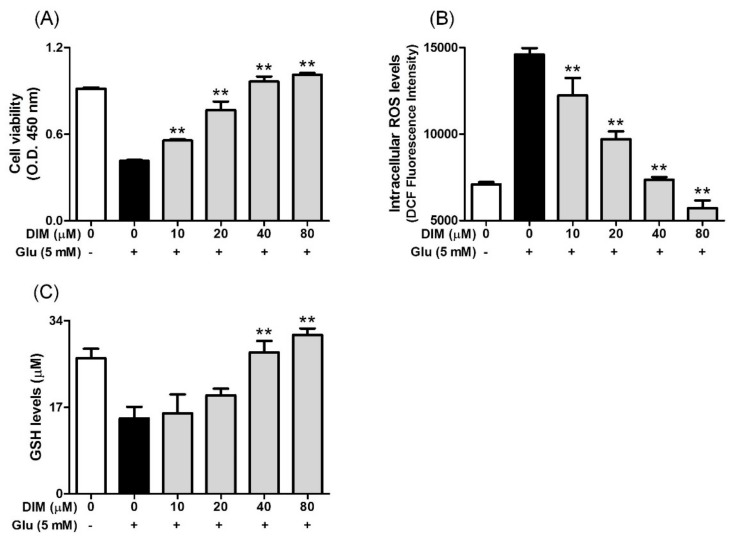
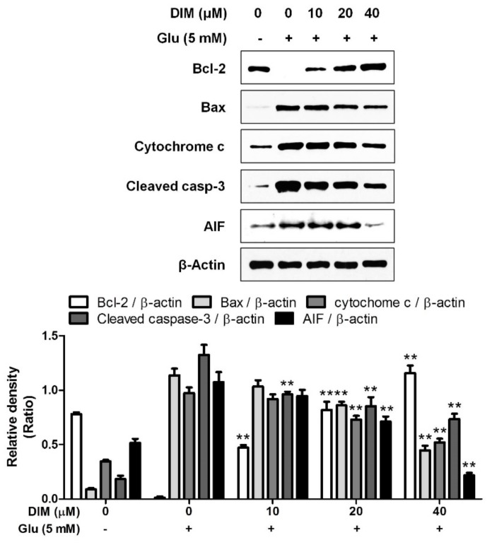
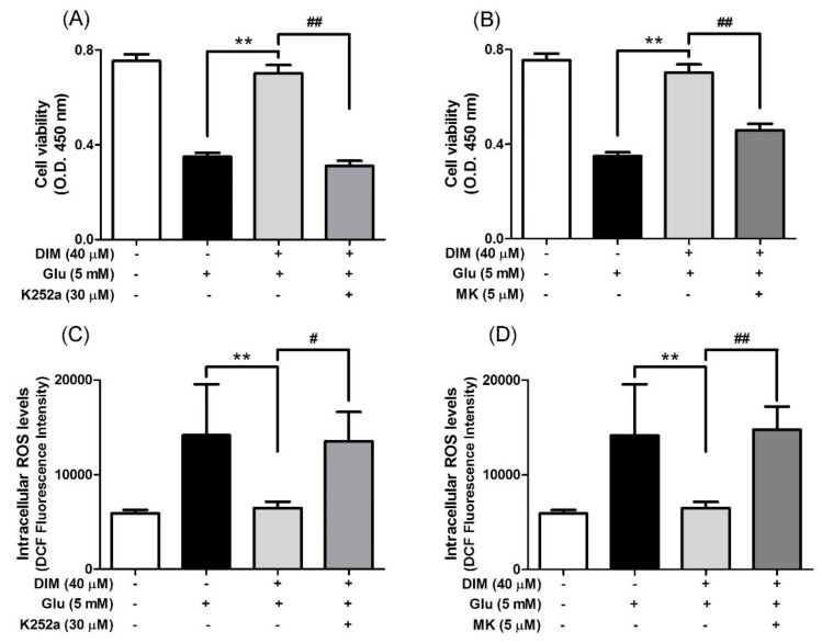
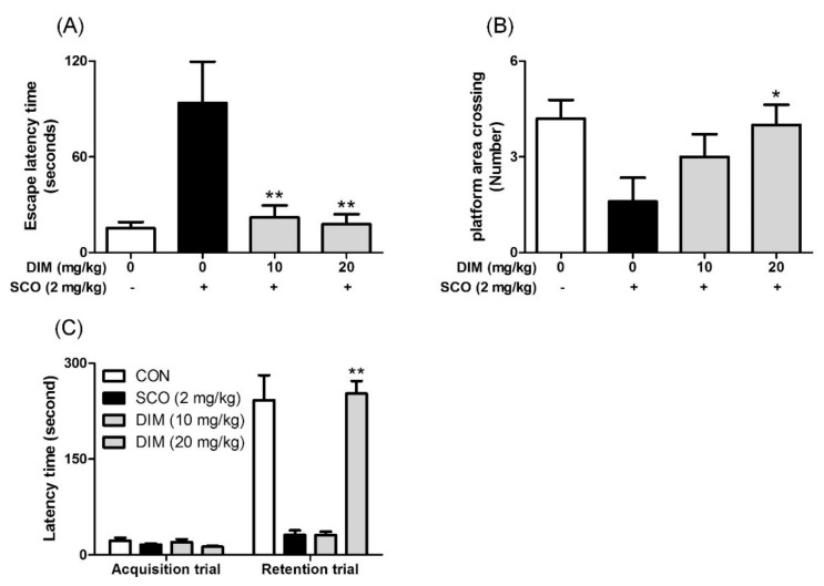
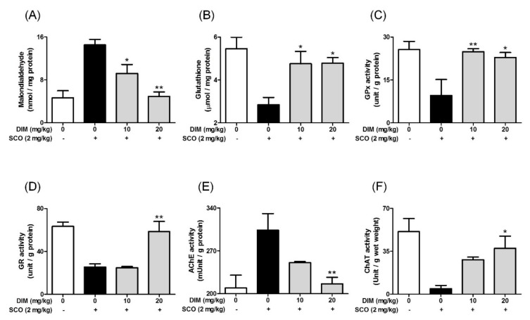
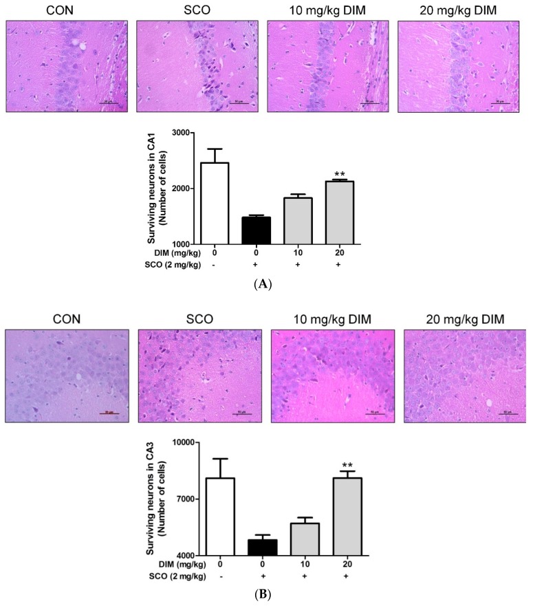
3,3'-Diindolylmethane (DIM), a metabolite of indole-3-carbinol present in Brassicaceae vegetables, possesses various health-promoting effects. Nonetheless, the effect of DIM on neurodegenerative diseases has not been elucidated clearly. In this study, we hypothesized DIM may protect neuronal cells against oxidative stress-induced apoptosis by promoting the formation of brain-derived neurotrophic factor (BDNF) and antioxidant enzymes through stabilizing the activation of the tropomyosin-related kinase receptor B (TrkB) cascade and we investigated the effect of DIM on oxidative stress-mediated neurodegenerative models. DIM protected neuronal cells against oxidative stress-induced apoptosis by regulating the expression of apoptosis-related proteins in glutamate-treated HT-22 cells. Additionally, DIM improved the expression of BDNF and antioxidant enzymes, such as heme oxygenase-1, glutamate-cysteine ligase catalytic subunit, and NAD(P)H quinine oxidoreductase-1, by promoting the activation of the TrkB/protein kinase B (Akt) pathway in the cells. Consistent with in vitro studies, DIM attenuated memory impairment by protecting hippocampal neuronal cells against oxidative damage in scopolamine-treated mice. Conclusionally, DIM exerted neuroprotective and antioxidant actions through the activation of both BDNF production and antioxidant enzyme formation in accordance with the TrkB/Akt pathway in neuronal cells. Such an effect of DIM may provide information for the application of DIM in the prevention of and therapy for neurodegenerative diseases.
Keywords: 3,3’-diindolylmethane; antioxidant enzymes; brain-derived neurotrophic factor; hippocampal neuronal cells; neurodegenerative disease.
Conflict of interest statement
The authors declare no conflict of interest.
Figures

Similar articles
-
Neuroprotective action of N-acetyl serotonin in oxidative stress-induced apoptosis through the activation of both TrkB/CREB/BDNF pathway and Akt/Nrf2/Antioxidant enzyme in neuronal cells.Redox Biol. 2017 Apr;11:592-599. doi: 10.1016/j.redox.2016.12.034. Epub 2017 Jan 6. Redox Biol. 2017. PMID: 28110215 Free PMC article.
-
Neuroprotective Effect of Carotenoid-Rich Enteromorpha prolifera Extract via TrkB/Akt Pathway against Oxidative Stress in Hippocampal Neuronal Cells.Mar Drugs. 2020 Jul 19;18(7):372. doi: 10.3390/md18070372. Mar Drugs. 2020. PMID: 32707633 Free PMC article.
-
Mulberry fruit improves memory in scopolamine-treated mice: role of cholinergic function, antioxidant system, and TrkB/Akt signaling.Nutr Neurosci. 2021 Dec;24(12):940-950. doi: 10.1080/1028415X.2019.1696613. Epub 2019 Dec 2. Nutr Neurosci. 2021. PMID: 31793392
-
Indole-3-Carbinol and Its Derivatives as Neuroprotective Modulators.Brain Sci. 2024 Jul 2;14(7):674. doi: 10.3390/brainsci14070674. Brain Sci. 2024. PMID: 39061415 Free PMC article. Review.
-
Neuroprotection Against Oxidative Stress: Phytochemicals Targeting TrkB Signaling and the Nrf2-ARE Antioxidant System.Front Mol Neurosci. 2020 Jul 2;13:116. doi: 10.3389/fnmol.2020.00116. eCollection 2020. Front Mol Neurosci. 2020. PMID: 32714148 Free PMC article. Review.
Cited by
-
Bee Venom Activates the Nrf2/HO-1 and TrkB/CREB/BDNF Pathways in Neuronal Cell Responses against Oxidative Stress Induced by Aβ1-42.Int J Mol Sci. 2022 Jan 21;23(3):1193. doi: 10.3390/ijms23031193. Int J Mol Sci. 2022. PMID: 35163115 Free PMC article.
-
Beneficial Health Effects of Glucosinolates-Derived Isothiocyanates on Cardiovascular and Neurodegenerative Diseases.Molecules. 2022 Jan 19;27(3):624. doi: 10.3390/molecules27030624. Molecules. 2022. PMID: 35163897 Free PMC article. Review.
-
3,3'-diindolylmethane inhibits LPS-induced human chondrocytes apoptosis and extracellular matrix degradation by activating PI3K-Akt-mTOR-mediated autophagy.Front Pharmacol. 2022 Nov 10;13:999851. doi: 10.3389/fphar.2022.999851. eCollection 2022. Front Pharmacol. 2022. PMID: 36438802 Free PMC article.
-
Neuroprotective Activity of Polyphenol-Rich Ribes diacanthum Pall against Oxidative Stress in Glutamate-Stimulated HT-22 Cells and a Scopolamine-Induced Amnesia Animal Model.Antioxidants (Basel). 2020 Sep 21;9(9):895. doi: 10.3390/antiox9090895. Antioxidants (Basel). 2020. PMID: 32967207 Free PMC article.
-
3,3'-Diindolylmethane Supplementation Maintains Oocyte Quality by Reducing Oxidative Stress and CEP-1/p53-Mediated Regulation of Germ Cells in a Reproductively Aged Caenorhabditis elegans Model.Antioxidants (Basel). 2022 May 11;11(5):950. doi: 10.3390/antiox11050950. Antioxidants (Basel). 2022. PMID: 35624814 Free PMC article.
References
-
- Fuentes F., Paredes-Gonzalez X., Kong A.N. Dietary glucosinolates sulforaphane, phenethyl isothiocyanate, indole-3-carbinol/3,3’-diindolylmethane: Anti-oxidative stress/inflammation, nrf2, epigenetics/epigenomics and in vivo cancer chemopreventive efficacy. Curr. Pharmacol. Rep. 2015;1:179–196. doi: 10.1007/s40495-015-0017-y. - DOI - PMC - PubMed
Grants and funding
LinkOut - more resources
Full Text Sources
