

Inhibition of MELK Protooncogene as an Innovative Treatment for Intrahepatic Cholangiocarcinoma
- PMID: 31861475
- PMCID: PMC7023300
- DOI: 10.3390/medicina56010001
Inhibition of MELK Protooncogene as an Innovative Treatment for Intrahepatic Cholangiocarcinoma
Abstract
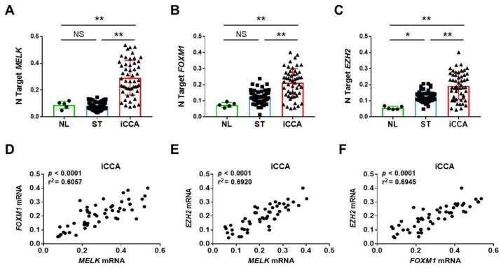
Background and Objectives: Intrahepatic cholangiocarcinoma (iCCA) is a pernicious tumor characterized by a dismal outcome and scarce therapeutic options. To substantially improve the prognosis of iCCA patients, a better understanding of the molecular mechanisms responsible for development and progression of this disease is imperative. In the present study, we aimed at elucidating the role of the maternal embryonic leucine zipper kinase (MELK) protooncogene in iCCA. Materials and Methods: We analyzed the expression of MELK and two putative targets, Forkhead Box M1 (FOXM1) and Enhancer of Zeste Homolog 2 (EZH2), in a collection of human iCCA by real-time RT-PCR and immunohistochemistry (IHC). The effects on iCCA growth of both the multi-kinase inhibitor OTSSP167 and specific small-interfering RNA (siRNA) against MELK were investigated in iCCA cell lines. Results: Expression of MELK was significantly higher in tumors than in corresponding non-neoplastic liver counterparts, with highest levels of MELK being associated with patients' shorter survival length. In vitro, OTSSP167 suppressed the growth of iCCA cell lines in a dose-dependent manner by reducing proliferation and inducing apoptosis. These effects were amplified when OTSSP167 administration was coupled to the DNA-damaging agent doxorubicin. Similar results, but less remarkable, were obtained when MELK was silenced by specific siRNA in the same cells. At the molecular level, siRNA against MELK triggered downregulation of MELK and its targets. Finally, we found that MELK is a downstream target of the E2F1 transcription factor. Conclusion: Our results indicate that MELK is ubiquitously overexpressed in iCCA, where it may represent a prognostic indicator and a therapeutic target. In particular, the combination of OTSSP167 (or other, more specific MELK inhibitors) with DNA-damaging agents might be a potentially effective therapy for human iCCA.
Keywords: EZH2; FOXM1; MELK; intrahepatic cholangiocarcinoma; targeted therapies.
Conflict of interest statement
The authors declare no conflicts of interest.
Figures


Similar articles
-
Inhibition of maternal embryonic leucine zipper kinase with OTSSP167 displays potent anti-leukemic effects in chronic lymphocytic leukemia.Oncogene. 2018 Oct;37(41):5520-5533. doi: 10.1038/s41388-018-0333-x. Epub 2018 Jun 12. Oncogene. 2018. PMID: 29895969
-
MELK-T1, a small-molecule inhibitor of protein kinase MELK, decreases DNA-damage tolerance in proliferating cancer cells.Biosci Rep. 2015 Oct 2;35(6):e00267. doi: 10.1042/BSR20150194. Biosci Rep. 2015. PMID: 26431963 Free PMC article.
-
Effective growth-suppressive activity of maternal embryonic leucine-zipper kinase (MELK) inhibitor against small cell lung cancer.Oncotarget. 2016 Mar 22;7(12):13621-33. doi: 10.18632/oncotarget.7297. Oncotarget. 2016. PMID: 26871945 Free PMC article.
-
Maternal embryonic leucine zipper kinase: key kinase for stem cell phenotype in glioma and other cancers.Mol Cancer Ther. 2014 Jun;13(6):1393-8. doi: 10.1158/1535-7163.MCT-13-0764. Epub 2014 May 2. Mol Cancer Ther. 2014. PMID: 24795222 Free PMC article. Review.
-
Enigmatic MELK: The controversy surrounding its complex role in cancer.J Biol Chem. 2020 Jun 12;295(24):8195-8203. doi: 10.1074/jbc.REV120.013433. Epub 2020 Apr 29. J Biol Chem. 2020. PMID: 32350113 Free PMC article. Review.
Cited by
-
Wnt/β-catenin signaling as an emerging potential key pharmacological target in cholangiocarcinoma.Biosci Rep. 2020 Mar 27;40(3):BSR20193353. doi: 10.1042/BSR20193353. Biosci Rep. 2020. PMID: 32140709 Free PMC article. Review.
-
Lactic acidosis induces metabolic and phenotypic reprogramming in cholangiocarcinoma cells via the upregulation of thrombospondin-1.Cancer Sci. 2023 Apr;114(4):1541-1555. doi: 10.1111/cas.15699. Epub 2023 Jan 28. Cancer Sci. 2023. PMID: 36562400 Free PMC article.
-
Cloning, tissue distribution, expression pattern, and function of porcine maternal embryonic leucine zipper kinase.Ann Transl Med. 2020 Mar;8(5):239. doi: 10.21037/atm.2020.03.46. Ann Transl Med. 2020. PMID: 32309386 Free PMC article.
-
Up-Regulation of MELK Promotes Cell Growth and Invasion by Accelerating G1/S Transition and Indicates Poor Prognosis in Lung Adenocarcinoma.Mol Biotechnol. 2025 Apr;67(4):1584-1596. doi: 10.1007/s12033-024-01143-4. Epub 2024 Apr 27. Mol Biotechnol. 2025. PMID: 38676754
References
-
- Banales J.M., Cardinale V., Carpino G., Marzioni M., Andersen J.B., Invernizzi P., Lind G.E., Folseraas T., Forbes S.J., Fouassier L., et al. Expert consensus document: Cholangiocarcinoma: Current knowledge and future perspectives consensus statement from the European Network for the Study of Cholangiocarcinoma (ENS-CCA) Nat. Rev. Gastroenterol. Hepatol. 2016;13:261–280. doi: 10.1038/nrgastro.2016.51. - DOI - PubMed
-
- Khan S.A., Davidson B.R., Goldin R.D., Heaton N., Karani J., Pereira S.P., Rosenberg W.M.C., Tait P., Taylor-Robinson S.D., Thillainayagam A.V., et al. Guidelines for the diagnosis and treatment of cholangiocarcinoma: An update. Gut. 2012;61:1657–1669. doi: 10.1136/gutjnl-2011-301748. - DOI - PubMed
MeSH terms
Substances
LinkOut - more resources
Full Text Sources
Miscellaneous
