. 2019 Dec 18;25(1):13.
doi: 10.3390/molecules25010013.

Inhibitory Effects of Myrtucommuacetalone 1 (MCA-1) from Myrtus communis on Inflammatory Response in Mouse Macrophages

Affiliations

Inhibitory Effects of Myrtucommuacetalone 1 (MCA-1) from Myrtus communis on Inflammatory Response in Mouse Macrophages

Samreen Soomro et al. Molecules. .

Abstract

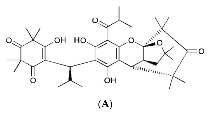
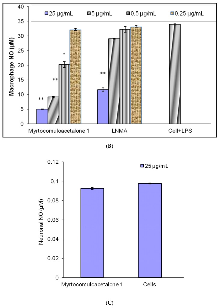
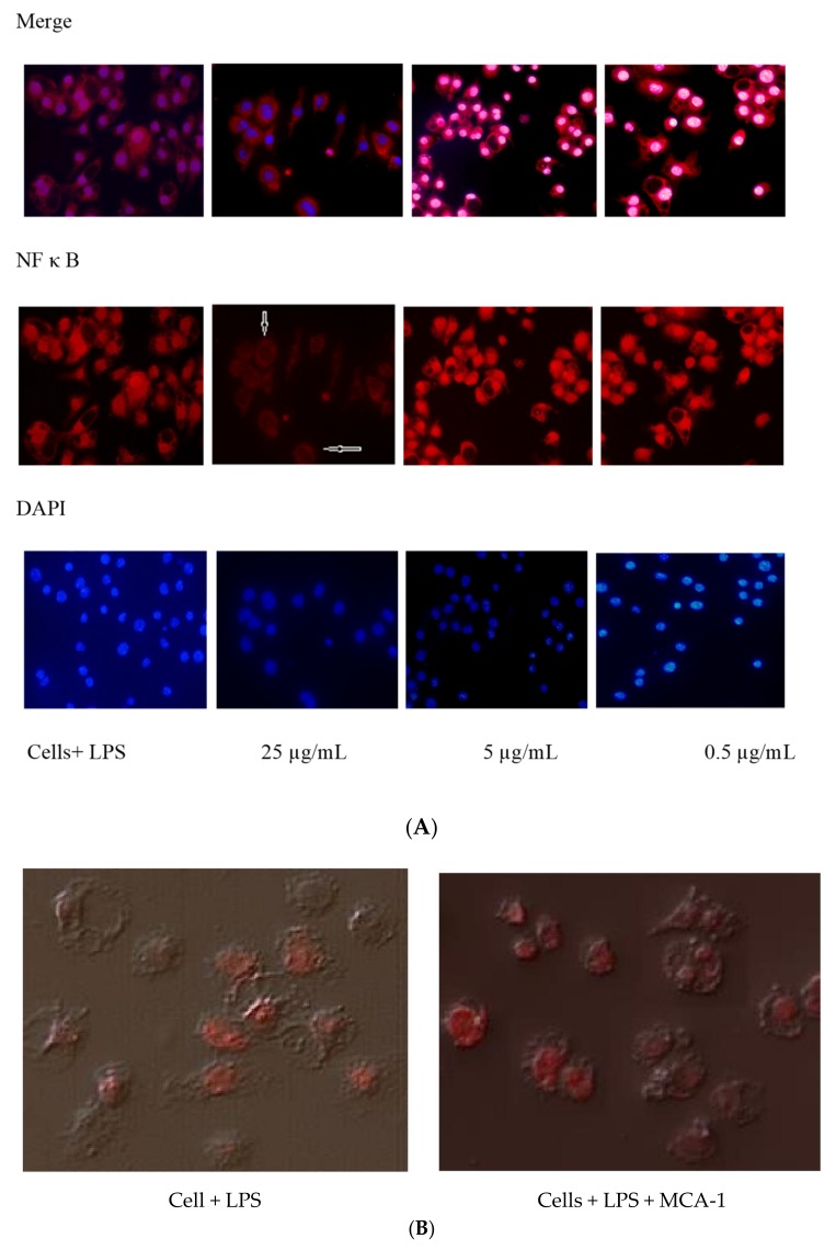
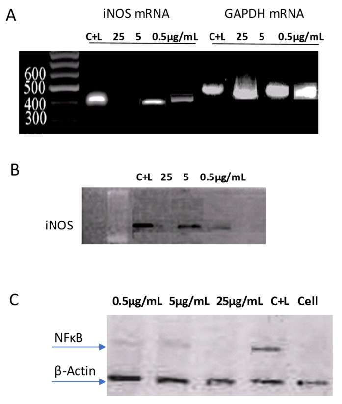
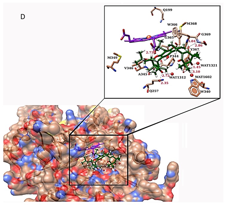
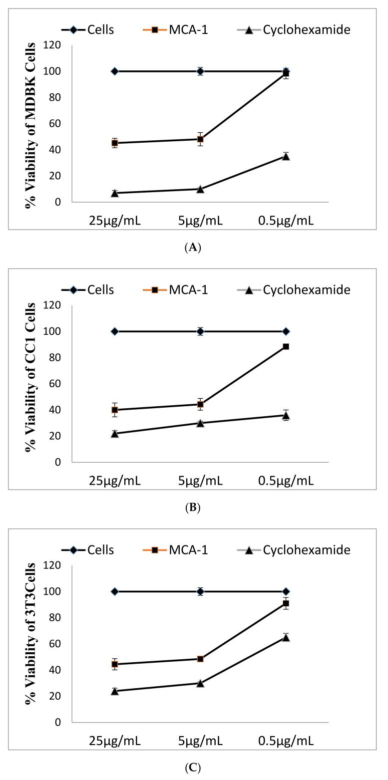
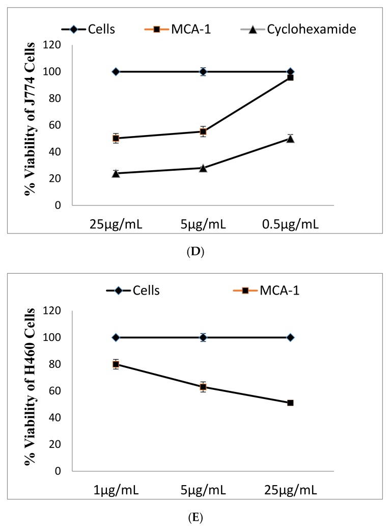
(1) Introduction: Reactive oxygen species (ROS) and nitric oxide (NO) are key signaling molecules that play important roles in the progression of inflammatory disorders. The objective of this study was to explore the use of myrtucommuacetalone-1 (MCA-1), as a novel compound of natural origin and a potential anti-inflammatory agent. (2) Methodology: The anti-inflammatory potential of MCA-1, which was isolated from Myrthus communis Linn, was determined by assaying superoxide, hydrogen peroxide, and nitric oxide production in macrophages. Furthermore, the effects of the compound were analyzed via phosphorylation and translocation of the transcription factor NF kappa B, which is a key regulator of iNOS activation. The effect of MCA-1 on the inducible nitric oxide synthase (iNOS) enzyme was also examined using in silico docking studies. The anticancer potential for MCA-1 was evaluated with an MTT cytotoxic assay. (3) Results: In stimulated macrophages, MCA-1 inhibited superoxide production by 48%, hydrogen peroxide by 53%, and nitric oxide (NO) with an IC50 of <1 µg/mL. MCA-1 also showed a very strong binding pattern within the active site of the inducible nitric oxide synthase enzyme. Furthermore, 25 µg/mL of MCA-1 inhibited inducible nitric oxide synthase expression and abolished transcription factor (NFκB) phosphorylation and translocation to the nucleus. Cytotoxicity analyses of MCA-1 on 3T3 mouse fibroblasts, CC1 liver cell line, J774.2, macrophages and MDBK bovine kidney epithelial cell, yielded IC50 values of 6.53 ± 1.2, 4.6 ± 0.7, 5 ± 0.8, and 4.6 ± 0.7, µg/mL, respectively. (4) Conclusion: Our results suggest that MCA-1, a major phloroglucinol-type compound, shows strong anti-inflammatory activity and has a potential to be a leading therapeutic agent in the future.

Keywords: inflammation; macrophages; nitric oxide; respiratory burst.


Conflict of interest statement

The authors declare no conflict of interest.

Figures

Figure 1
Figure 1
(A) Structure of myrtucommuacetalone 1 (MCA-1). (B) Effect of myrtucommuacetalone 1 (MCA-1) on nitric oxide (NO) release by activated THP1 monocytes. THP Cells (2 × 10⁵) were incubated in the presence (25, 5, 0.5, 0.25 µg/mL) or absence of the compound (control) for 48 h, and (C) the effects of the compound at 25 µg/mL on the basal level of neuronal NO in U 138MG neuronal cells were observed. The supernatant was analyzed for the presence of nitrite using the Griess method. Values are expressed as mean ± SD for triplicate experiments. The results were compared with NG Monomethyl L arginine (LNMA), a known inhibitor of NO. * p < 0.05, ** p < 0.005.
Figure 1
Figure 1
(A) Structure of myrtucommuacetalone 1 (MCA-1). (B) Effect of myrtucommuacetalone 1 (MCA-1) on nitric oxide (NO) release by activated THP1 monocytes. THP Cells (2 × 10⁵) were incubated in the presence (25, 5, 0.5, 0.25 µg/mL) or absence of the compound (control) for 48 h, and (C) the effects of the compound at 25 µg/mL on the basal level of neuronal NO in U 138MG neuronal cells were observed. The supernatant was analyzed for the presence of nitrite using the Griess method. Values are expressed as mean ± SD for triplicate experiments. The results were compared with NG Monomethyl L arginine (LNMA), a known inhibitor of NO. * p < 0.05, ** p < 0.005.
Figure 2
Figure 2
Lipopolysaccharide (LPS) induced nuclear translocation of the p65 subunit of NF kappa B and p38 kinase in J774 cells. The effects of MCA-1 at concentration 25 µg/mL (arrows) showed the absence of the NF kappa B transcription factor inside the nucleus at 25 µg/mL (A) and had no effect on p38 kinase translocation (B). The cells were examined at 20× magnification under the TRITC and DAPI channels using a Nikon TE-2000 epifluorescence microscope. The picture merge was performed using Adobe Photoshop.
Figure 3
Figure 3
(A) Effect of MCA-1 on iNOS expression. J774 macrophages treated with 30 µg/mL of LPS to induce iNOS expression and total RNA were extracted to determine iNOS expression using RT-PCR, as described in the text. (B) Effect of MCA-1 on iNOS Protein (C)Effect of MCA-1 on NFkB phosphorylation. J774 macrophages were treated with LPS in the presence of varying concentrations of MCA-1 for 1 h to induce NFκB phosphorylation and for 48 h to monitor iNOS expression. A total of 35 μg of protein from the cell lysate was resolved on an SDS-PAGE and proteins were detected using specific antibodies against phosphorylated NFkB or iNOS, as described in the text. Sample loading was monitored by detecting β-actin with specific antibodies. The blots shown are representatives of two independent experiments. (D) The 3D structure of murine iNOS and the binding mode of MCA-1 at the active site of iNOS is shown in the box. The active site residues are depicted by the coral sticks, the active site of heme is shown by the purple sticks, and the compound is shown by the green sticks.
Figure 3
Figure 3
(A) Effect of MCA-1 on iNOS expression. J774 macrophages treated with 30 µg/mL of LPS to induce iNOS expression and total RNA were extracted to determine iNOS expression using RT-PCR, as described in the text. (B) Effect of MCA-1 on iNOS Protein (C)Effect of MCA-1 on NFkB phosphorylation. J774 macrophages were treated with LPS in the presence of varying concentrations of MCA-1 for 1 h to induce NFκB phosphorylation and for 48 h to monitor iNOS expression. A total of 35 μg of protein from the cell lysate was resolved on an SDS-PAGE and proteins were detected using specific antibodies against phosphorylated NFkB or iNOS, as described in the text. Sample loading was monitored by detecting β-actin with specific antibodies. The blots shown are representatives of two independent experiments. (D) The 3D structure of murine iNOS and the binding mode of MCA-1 at the active site of iNOS is shown in the box. The active site residues are depicted by the coral sticks, the active site of heme is shown by the purple sticks, and the compound is shown by the green sticks.
Figure 4
Figure 4
Effect of MCA-1 on cellular toxicity. MDBK kidney (A), CC1 liver (B), 3T3 NIH mouse fibroblasts (C) and J774.2 mouse macrophage (D) cell lines were incubated in the absence (■) and presence (♦) of varying concentrations of MCA-1 for 48 h. The cytotoxic effects of MCA-1 were determined using the MTT assay, as described in the text. The effect of the cytotoxic drug cyclohexamide (▲) was also determined for comparison. (E) Anticancer activity of myrtocomuloaetalone1 on the H640 lung cancer cell line. H640 cells were incubated in the presence (■) or absence (♦) of varying concentrations of MCA-1 for 48 h. The cell viability was determined by FACS analysis, as described in the text. The values are represented as the mean ± SD for triplicate experiments. The data were analyzed by cell quest pro. Significant differences are reported at p < 0.05. The values are represented by the mean ± SD for triplicate experiments.
Figure 4
Figure 4
Effect of MCA-1 on cellular toxicity. MDBK kidney (A), CC1 liver (B), 3T3 NIH mouse fibroblasts (C) and J774.2 mouse macrophage (D) cell lines were incubated in the absence (■) and presence (♦) of varying concentrations of MCA-1 for 48 h. The cytotoxic effects of MCA-1 were determined using the MTT assay, as described in the text. The effect of the cytotoxic drug cyclohexamide (▲) was also determined for comparison. (E) Anticancer activity of myrtocomuloaetalone1 on the H640 lung cancer cell line. H640 cells were incubated in the presence (■) or absence (♦) of varying concentrations of MCA-1 for 48 h. The cell viability was determined by FACS analysis, as described in the text. The values are represented as the mean ± SD for triplicate experiments. The data were analyzed by cell quest pro. Significant differences are reported at p < 0.05. The values are represented by the mean ± SD for triplicate experiments.

Similar articles

Cited by

  • The therapeutic value of Myrtus communis L.: an updated review.
    Al-Snafi AE, Teibo JO, Shaheen HM, Akinfe OA, Teibo TKA, Emieseimokumo N, Elfiky MM, Al-Kuraishy HM, Al-Garbeeb AI, Alexiou A, Papadakis M, Mahana HAM, Younes AM, Elbanna OA, Qasem AAR, Shahin IYI, Batiha GE. Al-Snafi AE, et al. Naunyn Schmiedebergs Arch Pharmacol. 2024 Jul;397(7):4579-4600. doi: 10.1007/s00210-024-02958-3. Epub 2024 Feb 6. Naunyn Schmiedebergs Arch Pharmacol. 2024. PMID: 38319389 Free PMC article. Review.

References

    1. Semeraro N., Ammollo C.T., Semeraro F., Colucci M. Sepsis-associated dissemenated intravascular coagulation and thromboembolic disease. J. Hematol. Infect. Dis. 2010;2:2010–2024. - PMC - PubMed
    1. Choudhary M.I., Khan N., Ahmad M., Yousuf S., Fun H.K., Soomro S., Asif M., Mesaik M.A., Shaheen F. New inhibitors of ROS generation and T-cell proliferation from Myrtus communis. Org. Lett. 2013;15:1862–1865. doi: 10.1021/ol4002976. - DOI - PubMed
    1. Aktan F. iNOS-mediated nitric oxide production and its regulation. Life Sci. 2004;75:639–653. doi: 10.1016/j.lfs.2003.10.042. - DOI - PubMed
    1. Macdonald J., Galley H.F., Webster N.R. Oxidative stress and gene expression in sepsis. Br. J. Anaesth. 2003;90:221–232. doi: 10.1093/bja/aeg034. - DOI - PubMed
    1. Karin M. NF-κB as a critical link between inflammation and cancer. Cold Spring Harb Perspect Biol. 2009;1:5. doi: 10.1101/cshperspect.a000141. - DOI - PMC - PubMed

MeSH terms

LinkOut - more resources