. 2019 Dec 18;10(1):8.
doi: 10.3390/biom10010008.

Functional Analysis of DNMT3A DNA Methyltransferase Mutations Reported in Patients with Acute Myeloid Leukemia

Affiliations

Functional Analysis of DNMT3A DNA Methyltransferase Mutations Reported in Patients with Acute Myeloid Leukemia

Daria A Khrabrova et al. Biomolecules. .

Abstract

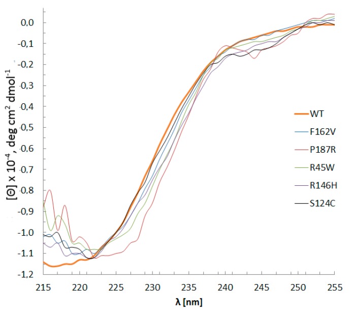
In mammals, DNA methylation is necessary for the maintenance of genomic stability, gene expression regulation, and other processes. During malignant diseases progression, changes in both DNA methylation patterns and DNA methyltransferase (MTase) genes are observed. Human de novo MTase DNMT3A is most frequently mutated in acute myeloid leukemia (AML) with a striking prevalence of R882H mutation, which has been extensively studied. Here, we investigate the functional role of the missense mutations (S714C, R635W, R736H, R771L, P777R, and F752V) found in the catalytic domain of DNMT3A in AML patients. These were accordingly mutated in the murine Dnmt3a catalytic domain (S124C, R45W, R146H, R181L, P187R, and F162V) and in addition, one-site CpG-containing DNA substrates were used as a model system. The 3-15-fold decrease (S124C and P187R) or complete loss (F162V, R45W, and R146H) of Dnmt3a-CD methylation activity was observed. Remarkably, Pro 187 and Arg 146 are not located at or near the Dnmt3a functional motives. Regulatory protein Dnmt3L did not enhance the methylation activity of R45W, R146H, P187R, and F162V mutants. The key steps of the Dnmt3a-mediated methylation mechanism, including DNA binding and transient covalent intermediate formation, were examined. There was a complete loss of DNA-binding affinity for R45W located in the AdoMet binding region and for R146H. Dnmt3a mutants studied in vitro suggest functional impairment of DNMT3A during pathogenesis.

Keywords: DNA methylation; DNA methyltransferase Dnmt3a; DNA-protein binding; S-adenosyl-L-methionine; leukemia; missense mutations.


Conflict of interest statement

Authors declare no conflict of interest in financial or any other sphere.

Figures

Figure A1
Figure A1
R181L Western blot. Lane 1 represents the protein ladder; Lanes 2 and 3 represent protein dilution 1:10 and 1:15, respectively. Fifteen percent PAG with 10% SDS, 1:5000 mouse HRP monoclonal anti-His6-tag antibodies. Molecular mass of the Dnmt3a-CD 37 kDa, the expected second lane of 20 kDa is not observed.
Figure 1
Figure 1
DNMT3A expression during different cancer types among patients (created with CBioPortal tools based on The Cancer Genome Atlas (TCGA) database information). The height of the column corresponds to the change in mRNA production. Red dots mark point mutations, blue dots mark no mutation, white dots mark not sequenced samples, red triangles mark frameshifts and splicing disorders, red squares mark other types of disorders. One dot represents one blood sample.
Figure 2
Figure 2
Alignment of the sequences of the catalytic domains of human and murine methyltransferases (MTases) Dnmt3a and Dnmt3b and prokaryotic MTases HhaI and HaeIII with underlined conserved motives (I-X) [33] and location of mutated amino acid residues. The negatively charged amino acids are given in red, the positively charged ones are given in blue, the polar ones are given in violet, the aromatic/non-polar ones are given in green, proline is given in gray, cysteine is given in wine-red, glycine is given in light orange, histidine is given in magenta. Highly conserved amino acids are given stars above, conserved amino acids are given two dots above, often preserved amino acids are given one dot above. The mutated amino acid residues are indicated.
Figure 3
Figure 3
WT Dnmt3a-CD and mutants CD spectra. Buffer A with 0.1 mM AdoHcy, path length 0.5 mm, 15 °C, protein concentration 0.1–0.4 g/L. Data represents the averaged results from three measurements.
Figure 4
Figure 4
Effect of cancer-associated mutations of Dnmt3a-CD on DNA methylation. (a,b) Cleavage of 0.3 μM fCG/GCf with Hin6I endonuclease after its methylation with 2 μM WT Dnmt3a-CD or P187R; 20% PAG with 7 M urea; (c) Time courses of methylation of fCG/GCf by WT and mutant Dnmt3a-CD. [(CH3)-DNA] values were calculated based on the data from (a) and (b) (see Materials and Methods). t represents the time of methylation reaction.
Figure 5
Figure 5
Methylation by WT and mutant Dnmt3a-CD in the presence or absence of Dnmt3L during 2 h. (a) Reaction mixtures contained 0.3 μM fCG/GCf DNA substrate, 2 μM Dnmt3a-CD or mutant, and 25 μM AdoMet; data represent the averaged results from at least six independent experiments; (b) Reaction mixtures contained 120 nM fCG/GCf DNA substrate, 1 μM WT or mutant Dnmt3a-CD, and 1 μM Dnmt3L; data represent the averaged results from three independent experiments.
Figure 6
Figure 6
Binding curves for Dnmt3a-CD WT and mutants obtained upon titration of the fCG/CG DNA substrate (10 nM) in the presence of 0.1 mM AdoHcy. P represents the fluorescence polarization. The inset shows binding curves for P187R and F162V on a different scale. Data represent the averaged results from at least two independent experiments.
Figure 7
Figure 7
Analysis of the covalent complex formation of WT Dnmt3a-CD and mutants with 2-pyrimidinone-substituted DNA duplex fCG/GZ. Six micromolar WT Dnmt3a-CD or S124C, 0.2 μM fCG/GZ DNA substrate, 0.1 mM AdoHcy. Twelve percent PAG with 0.1% SDS. Lane 1 represents fCG/GZ, lanes 2 and 3 represent fCG/GZ with WT or S124C, respectively.
Figure 8
Figure 8
The fragment of DNA-(DNMT3A-CD)-DNMT3L complex structure (PDB: 6F57). DNA is given in orange sticks, DNMT3A-CD is colored by secondary structure (α-helix in green, β-sheet in blue, loops in white), the catalytic loop is colored in yellow, target recognition domain (TRD) is colored in magenta. Residues of interest within human DNMT3A-CD are numbered in accordance with murine Dnmt3a-CD and given in red.

References

    1. Bird A. DNA methylation patterns and epigenetic memory. Genes Dev. 2002;16:6–21. doi: 10.1101/gad.947102. - DOI - PubMed
    1. Deaton A.M., Bird A. CpG islands and the regulation of transcription. Genes Dev. 2011;25:1010–1022. doi: 10.1101/gad.2037511. - DOI - PMC - PubMed
    1. Jurkowska R.Z., Jurkowski T.P., Jeltsch A. Structure and function of mammalian DNA methyltransferases. Chembiochem. 2011;12:206–222. doi: 10.1002/cbic.201000195. - DOI - PubMed
    1. Hamidi T., Singh A.K., Chen T. Genetic alterations of DNA methylation machinery in human diseases. Epigenomics. 2015;7:247–265. doi: 10.2217/epi.14.80. - DOI - PubMed
    1. Robertson K.D. DNA methylation and human disease. Nat. Rev. Genet. 2005;6:597–610. doi: 10.1038/nrg1655. - DOI - PubMed

Publication types

MeSH terms

Substances