. 2019 Dec 19;9(1):8.
doi: 10.3390/jcm9010008.

Lipophilicity of Bacteriochlorin-Based Photosensitizers as a Determinant for PDT Optimization through the Modulation of the Inflammatory Mediators

Affiliations

Lipophilicity of Bacteriochlorin-Based Photosensitizers as a Determinant for PDT Optimization through the Modulation of the Inflammatory Mediators

Barbara Pucelik et al. J Clin Med. .

Abstract

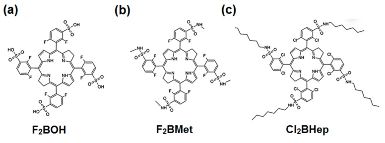
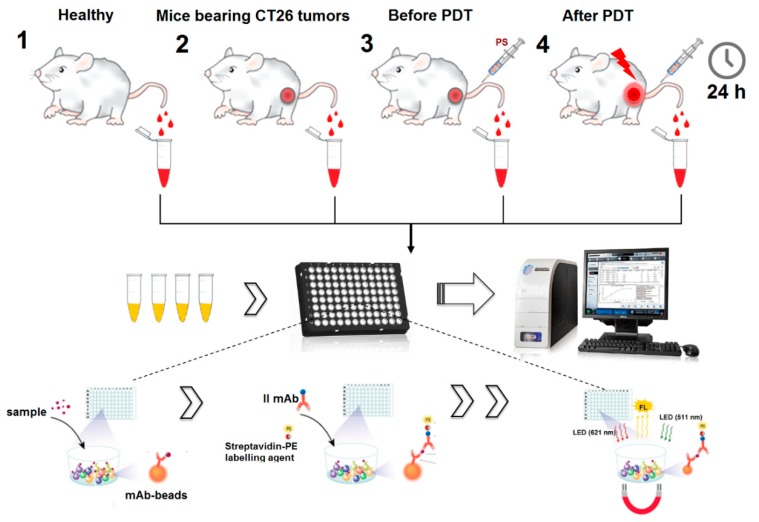
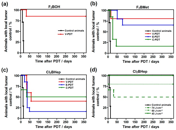
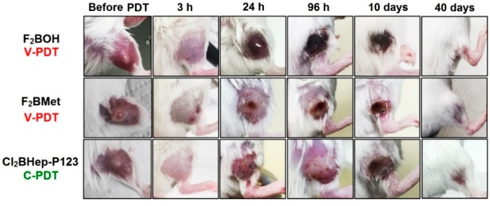
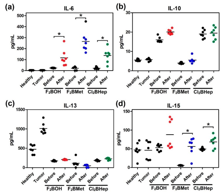
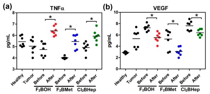
: Photodynamic therapy (PDT) augments the host antitumor immune response, but the role of the PDT effect on the tumor microenvironment in dependence on the type of photosensitizer and/or therapeutic protocols has not been clearly elucidated. We employed three bacteriochlorins (F2BOH, F2BMet and Cl2BHep) of different polarity that absorb near-infrared light (NIR) and generated a large amount of reactive oxygen species (ROS) to compare the PDT efficacy after various drug-to-light intervals: 15 min. (V-PDT), 3h (E-PDT) and 72h (C-PDT). We also performed the analysis of the molecular mechanisms of PDT crucial for the generation of the long-lasting antitumor immune response. PDT-induced damage affected the integrity of the host tissue and developed acute (protocol-dependent) local inflammation, which in turn led to the infiltration of neutrophils and macrophages. In order to further confirm this hypothesis, a number of proteins in the plasma of PDT-treated mice were identified. Among a wide range of cytokines (IL-6, IL-10, IL-13, IL-15, TNF-α, GM-CSF), chemokines (KC, MCP-1, MIP1α, MIP1β, MIP2) and growth factors (VEGF) released after PDT, an important role was assigned to IL-6. PDT protocols optimized for studied bacteriochlorins led to a significant increase in the survival rate of BALB/c mice bearing CT26 tumors, but each photosensitizer (PS) was more or less potent, depending on the applied DLI (15 min, 3 h or 72 h). Hydrophilic (F2BOH) and amphiphilic (F2BMet) PSs were equally effective in V-PDT (>80 cure rate). F2BMet was the most efficient in E-PDT (DLI = 3h), leading to a cure of 65 % of the animals. Finally, the most powerful PS in the C-PDT (DLI = 72 h) regimen turned out to be the most hydrophobic compound (Cl2BHep), allowing 100 % of treated animals to be cured at a light dose of only 45 J/cm2.

Keywords: bacteriochlorins; chemokines; cytokines; immune response.; photodynamic therapy.


Conflict of interest statement

The authors declare no conflict of interest.

Figures

Scheme 1
Scheme 1
Chemical structure of investigated halogenated bacteriochlorin derivatives.
Scheme 2
Scheme 2
Scheme for carrying out immunological analyses using the MAGPIX system.
Figure 1
Figure 1
Representative, normalized absorption and fluorescence spectra of F2BMet in dimethyl sulfoxide (DMSO).
Figure 2
Figure 2
Generation of reactive oxygen species (ROSs) in bacteriochlorin solution (in phosphate-buffered saline (PBS), dimethyl sulfoxide (DMSO) did not exceed 0.5%) assessed by the fluorescence probes selective towards: (a) overall radical species (APF), (b) hydroxyl radicals (HPF), (c) superoxide radical anion (DHE) or (d) SOSG specific for singlet oxygen. The concentrations of the probes were initially 10 μM, and bacteriochlorin was 5 μM in each sample. Samples were irradiated with a 735 ± 20 nm LED light. Data are presented as mean ± the standard error of the mean (SEM) (N = 12).
Figure 3
Figure 3
Photostability of sulfonated and sulfonamide bacteriochlorins in PBS solutions (<0.5% DMSO) under a near-infrared (NIR) light-emitting diode (LED) irradiation: (a) the photodegradation rate; (b) the changes in absorption spectra of F2BOH during irradiation.
Figure 4
Figure 4
The impact of Cl2BHep encapsulation in P123 micelles: (a) Absorption spectra of Cl2BHep registered in PBS (< 0.5% DMSO) and micellar solution in PBS after its encapsulation in Pluronic P123; (b) particle size distribution of Cl2BHep-P123 measured by dynamic light scattering (DLS).
Figure 5
Figure 5
In vitro activity of investigated photosensitizers: (a) time-dependent uptake of halogenated bacteriochlorins at 5 μM by CT26 cells determined based on fluorescence measurements of cellular extracts; (b) cytotoxicity in the dark toward CT26 cells determined by MTT assay after optimal incubation time estimated in uptake studies; (c) photodynamic efficacy of F2BOH, F2BMet and Cl2BHep after their incubation at 5 μM with CT26 for optimal time and irradiation with 735 ± 20 nm LED light; (d) phototoxicity of F2BOH at 5 μM without washing the medium before illumination, and of Cl2BHep encapsulated in P123 micelles at 5 μM. Data are presented as mean ± SEM (N = 12), * p < 0.05.
Figure 6
Figure 6
Confocal images of CT26 cells showing intracellular localization of Cl2BHep delivered in P123 micelles. Cells were marked with specific probes for mitochondria (Mito-Tracker), lysosomes (Lyso-Tracker) and endoplasmic reticulum (ER-Tracker).
Figure 7
Figure 7
Colocalization of the photosensitizer (PS) and organelle-specific probes (a) MitoTracker, (b) Lysotracker and (c) ERTracker determined and quantified based on the confocal microscopy images and analyzed by ZenSoftware (Zeiss, Jena, Germany).
Figure 8
Figure 8
Biodistribution of Cl2BHep encapsulated in P123 micelles in organs and tissues extracted from BALB/c mice bearing CT26 tumors determined at various periods of time after the injection. The data are presented as mean ± SEM (N = 6).
Figure 9
Figure 9
Kaplan–Meier curves of CT26 tumor regrowth after PDT treatment with: (a) F2BOH V-PDT; (b) F2BMet V-PDT, E-PDT and C-PDT; (c) Cl2BHep V-PDT, E-PDT and C-PDT; in all cases the tumor-bearing mice (N = 6–7) were irradiated with 74 J/cm2 dose of NIR laser light (130 mW, 748 nm) at 15 min, 3h or 72 h post-IV injection of 1.5 mg/kg drug dose. (d) presents the surviving data obtained for Cl2BHep-C-PDT after application of different laser light doses (light-dose de-escalation study) and 1.5 mg/kg drug dose.
Figure 10
Figure 10
The differences in the inflammatory reaction occurring in BALB/c mice bearing CT26 tumors before and after PDT using optimal protocols with F2BOH, F2BMet, and Cl2BHep as the photosensitizer.
Figure 11
Figure 11
Concentrations of selected interleukins: (a) IL-6; (b) IL-10; (c) IL-13; (d) IL-15 in the serum of mice before and after optimal PDT protocol with F2BOH (V-PDT), F2BMet (V-PDT) and Cl2BHep (C-PDT) as well as healthy mice and mice bearing CT26 tumor controls. Luminex, N = 6–7; each dot represents one individual, line represents a mean; * p < 0.05.
Figure 12
Figure 12
Concentrations of selected neutrophil chemokine attractants: (a) KC; (b) MIP-2; in the serum of mice before and after optimal PDT protocol with F2BOH (V-PDT), F2BMet (V-PDT) and Cl2BHep (C-PDT), as well as healthy mice and mice bearing CT26 tumor controls. Luminex, N = 6–7; each dot represents one individual, line represents a mean; * p < 0.05.
Figure 13
Figure 13
Concentrations of (a) TNFα; (b) VEGF in the serum of mice before and after optimal PDT protocol with F2BOH (V-PDT), F2BMet (V-PDT) and Cl2BHep (C-PDT) as well as healthy mice and mice bearing CT26 tumor controls. Luminex, N = 6–7; each dot represents one individual, line represents a mean; * p < 0.05.

Similar articles

Cited by

References

    1. Kessel D. Photodynamic Therapy: A Brief History. J. Clin. Med. 2019;8:1581. doi: 10.3390/jcm8101581. - DOI - PMC - PubMed
    1. Dabrowski J.M., Arnaut L.G. Photodynamic therapy (PDT) of cancer: From local to systemic treatment. Photochem. Photobiol. Sci. 2015;14:1765–1780. doi: 10.1039/C5PP00132C. - DOI - PubMed
    1. Dąbrowski J.M. Advances in Inorganic Chemistry. Volume 70. Elsevier; Amsterdam, The Netherlands: 2017. Reactive oxygen species in photodynamic therapy: Mechanisms of their generation and potentiation; pp. 343–394.
    1. Agostinis P., Berg K., Cengel K.A., Foster T.H., Girotti A.W., Gollnick S.O., Hahn S.M., Hamblin M.R., Juzeniene A., Kessel D., et al. Photodynamic therapy of cancer: An update. CA Cancer J. Clin. 2011;61:250–281. doi: 10.3322/caac.20114. - DOI - PMC - PubMed
    1. Krzykawska-Serda M., Dąbrowski J.M., Arnaut L.G., Szczygieł M., Urbańska K., Stochel G., Elas M. The role of strong hypoxia in tumors after treatment in the outcome of bacteriochlorin-based photodynamic therapy. Free Radic. Biol. Med. 2014;73:239–251. doi: 10.1016/j.freeradbiomed.2014.05.003. - DOI - PubMed

LinkOut - more resources