Review
. 2019 Dec 19;9(1):6.
doi: 10.3390/pathogens9010006.

The Role of Pseudomonas aeruginosa Lipopolysaccharide in Bacterial Pathogenesis and Physiology

Affiliations
Review

The Role of Pseudomonas aeruginosa Lipopolysaccharide in Bacterial Pathogenesis and Physiology

Steven M Huszczynski et al. Pathogens. .

Abstract

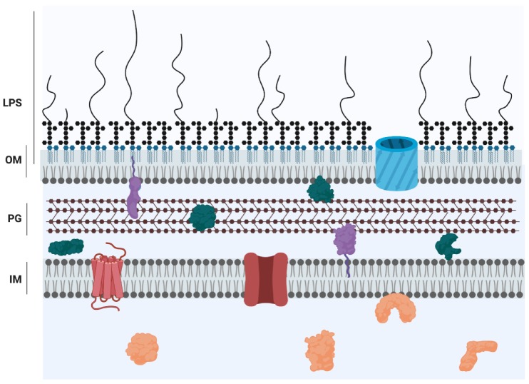
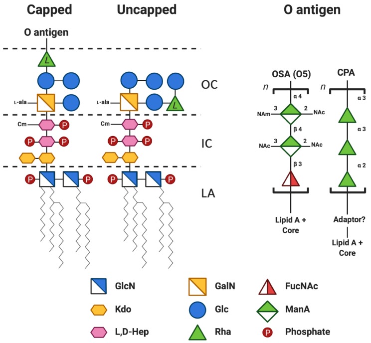
The major constituent of the outer membrane of Gram-negative bacteria is lipopolysaccharide (LPS), which is comprised of lipid A, core oligosaccharide, and O antigen, which is a long polysaccharide chain extending into the extracellular environment. Due to the localization of LPS, it is a key molecule on the bacterial cell wall that is recognized by the host to deploy an immune defence in order to neutralize invading pathogens. However, LPS also promotes bacterial survival in a host environment by protecting the bacteria from these threats. This review explores the relationship between the different LPS glycoforms of the opportunistic pathogen Pseudomonas aeruginosa and the ability of this organism to cause persistent infections, especially in the genetic disease cystic fibrosis. We also discuss the role of LPS in facilitating biofilm formation, antibiotic resistance, and how LPS may be targeted by new antimicrobial therapies.

Keywords: O antigen; antimicrobial resistance; biofilms; cystic fibrosis; host–pathogen interactions; lipopolysaccharide; pyocin.


Conflict of interest statement

The authors declare no conflict of interest. The funders had no role in the design of the study; in the collection, analyses, or interpretation of data; in the writing of the manuscript, or in the decision to publish the results.

Figures

Figure 1
Figure 1
Cartoon representation of the Gram-negative cell envelope. The inner membrane (IM) is a symmetric bilayer comprised of phospholipids while the outer membrane (OM) is an asymmetric bilayer containing phospholipids in the inner leaflet and LPS in the outer leaflet. The domains of LPS are represented as follows: lipid A, blue; core, black hexagons; O antigen, curved lines. The membranes are separated by the periplasmic space, which contains a thin layer of peptidoglycan (PG) [8]. Proteins are localized to all compartments and represented by the following colours: orange, cytoplasmic proteins; red, inner membrane proteins; purple, lipoproteins; green, periplasmic proteins; blue, outer membrane protein.
Figure 2
Figure 2
Simplified chemical structure of P. aeruginosa PAO1 (serotype O5) lipopolysaccharide. The structure is adapted from several studies [12,13,14,15,16,17] and coloured according to the Symbol Nomenclature for Glycans (SNFG) [18,19]. A more detailed review of the chemical structure of P. aeruginosa LPS can be found in [17]. The lipid A-core region can be capped (or not) with a variable number of O antigen repeats. The predominant penta-acylated lipid A structure is shown. For clarity, the following modifications to the core sugars are not shown: the phosphorylation sites on the two heptose residues are depicted as monophosphorylated but may contain mono- di- or triphosphates; non-stoichiometric O-acetylation of the outer core sugars; the phosphate at position 2 in Hep II is non-stoichiometrically modified with phosphoethanolamine. The l-configuration of the rhamnose in the core is denoted by l to distinguish it from d-rhamnose found in the CPA repeat unit. A short sugar adapter may be present between CPA and the lipid A-core. OC, outer core; IC, inner core; LA, lipid A; GlcN, glucosamine; GalN, galactosamine; FucNAc, N-acetyl-d-fucosamine Kdo, 3-deoxy-d-manno-oct-2-ulosonic acid; Glc, glucose; ManA, manuronic acid; l,d-Hep, l-glycero-d-manno-heptose; Rha, rhamnose; Cm, 7-O-carbamoylation; l-Ala, 2-l-alanylation; n, variable number of repeats; NAm, N-amidino; NAc, N-acetyl.
Figure 3
Figure 3
Summary of relevant P. aeruginosa O antigen, core, and lipid A interactions with antimicrobials and host defences. Arrows indicate binding of, or activation by, a specific LPS region while the flat-headed arrows indicate inhibition. The O antigen is coloured red, the core is coloured yellow, and lipid A is coloured blue.

References

    1. Rice L.B. Federal funding for the study of antimicrobial resistance in nosocomial pathogens: No ESKAPE. J. Infect. Dis. 2008;197:1079–1081. doi: 10.1086/533452. - DOI - PubMed
    1. Bedard E., Prevost M., Deziel E. Pseudomonas aeruginosa in premise plumbing of large buildings. Microbiologyopen. 2016;5:937–956. doi: 10.1002/mbo3.391. - DOI - PMC - PubMed
    1. Gellatly S.L., Hancock R.E.W. Pseudomonas aeruginosa: New insights into pathogenesis and host defenses. Pathog. Dis. 2013;67:159–173. doi: 10.1111/2049-632X.12033. - DOI - PubMed
    1. Rahme L.G., Stevens E.J., Wolfort S.F., Shao J., Tompkins R.G., Ausubel F.M. Common virulence factors for bacterial pathogenicity in plants and animals. Science. 1995;268:1899–1902. doi: 10.1126/science.7604262. - DOI - PubMed
    1. Schroth M.N., Cho J.J., Green S.K., Kominos S.D., Publishing M.S. Epidemiology of Pseudomonas aeruginosa in agricultural areas. J. Med. Microbiol. 2018;67:1191–1201. doi: 10.1099/jmm.0.000758. - DOI - PubMed

LinkOut - more resources