

Metabolic Hallmarks of Hepatic Stellate Cells in Liver Fibrosis
- PMID: 31861818
- PMCID: PMC7016711
- DOI: 10.3390/cells9010024
Metabolic Hallmarks of Hepatic Stellate Cells in Liver Fibrosis
Abstract
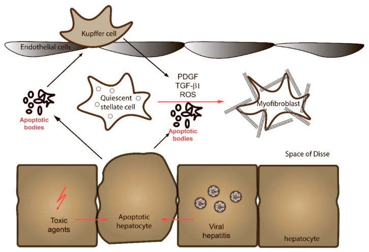
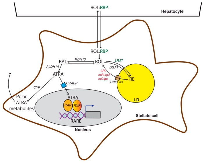
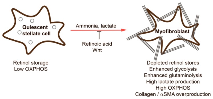
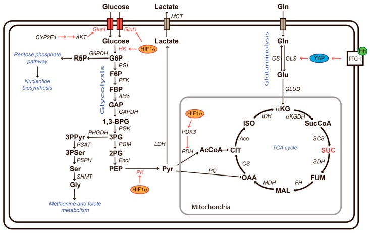
Liver fibrosis is a regenerative process that occurs after injury. It is characterized by the deposition of connective tissue by specialized fibroblasts and concomitant proliferative responses. Chronic damage that stimulates fibrogenic processes in the long-term may result in the deposition of excess matrix tissue and impairment of liver functions. End-stage fibrosis is referred to as cirrhosis and predisposes strongly to the loss of liver functions (decompensation) and hepatocellular carcinoma. Liver fibrosis is a pathology common to a number of different chronic liver diseases, including alcoholic liver disease, non-alcoholic fatty liver disease, and viral hepatitis. The predominant cell type responsible for fibrogenesis is hepatic stellate cells (HSCs). In response to inflammatory stimuli or hepatocyte death, HSCs undergo trans-differentiation to myofibroblast-like cells. Recent evidence shows that metabolic alterations in HSCs are important for the trans-differentiation process and thus offer new possibilities for therapeutic interventions. The aim of this review is to summarize current knowledge of the metabolic changes that occur during HSC activation with a particular focus on the retinol and lipid metabolism, the central carbon metabolism, and associated redox or stress-related signaling pathways.
Keywords: hepatic stellate cell; liver fibrosis; metabolism; trans-differentiation.
Conflict of interest statement
The authors declare no conflict of interest.
Figures
Similar articles
-
Coordinated signaling of activating transcription factor 6α and inositol-requiring enzyme 1α regulates hepatic stellate cell-mediated fibrogenesis in mice.Am J Physiol Gastrointest Liver Physiol. 2021 May 1;320(5):G864-G879. doi: 10.1152/ajpgi.00453.2020. Epub 2021 Mar 17. Am J Physiol Gastrointest Liver Physiol. 2021. PMID: 33728997 Free PMC article.
-
Hedgehog-YAP Signaling Pathway Regulates Glutaminolysis to Control Activation of Hepatic Stellate Cells.Gastroenterology. 2018 Apr;154(5):1465-1479.e13. doi: 10.1053/j.gastro.2017.12.022. Epub 2018 Jan 3. Gastroenterology. 2018. PMID: 29305935 Free PMC article.
-
Hepatic stellate cells role in the course of metabolic disorders development - A molecular overview.Pharmacol Res. 2021 Aug;170:105739. doi: 10.1016/j.phrs.2021.105739. Epub 2021 Jun 23. Pharmacol Res. 2021. PMID: 34171492 Review.
-
Wnt signaling in liver fibrosis: progress, challenges and potential directions.Biochimie. 2013 Dec;95(12):2326-35. doi: 10.1016/j.biochi.2013.09.003. Epub 2013 Sep 13. Biochimie. 2013. PMID: 24036368 Review.
-
Histone H3K27 methyltransferase EZH2 and demethylase JMJD3 regulate hepatic stellate cells activation and liver fibrosis.Theranostics. 2021 Jan 1;11(1):361-378. doi: 10.7150/thno.46360. eCollection 2021. Theranostics. 2021. PMID: 33391480 Free PMC article.
Cited by
-
Blockade of 11β-hydroxysteroid dehydrogenase type 1 ameliorates metabolic dysfunction-associated steatotic liver disease and fibrosis.Heliyon. 2024 Oct 18;10(20):e39534. doi: 10.1016/j.heliyon.2024.e39534. eCollection 2024 Oct 30. Heliyon. 2024. PMID: 39498052 Free PMC article.
-
Complexation with Random Methyl-β-Cyclodextrin and (2-Hidroxypropyl)-β-Cyclodextrin Enhances In Vivo Anti-Fibrotic and Anti-Inflammatory Effects of Chrysin via the Inhibition of NF-κB and TGF-β1/Smad Signaling Pathways and Modulation of Hepatic Pro/Anti-Fibrotic miRNA.Int J Mol Sci. 2021 Feb 13;22(4):1869. doi: 10.3390/ijms22041869. Int J Mol Sci. 2021. PMID: 33668543 Free PMC article.
-
NAFLD: Mechanisms, Treatments, and Biomarkers.Biomolecules. 2022 Jun 13;12(6):824. doi: 10.3390/biom12060824. Biomolecules. 2022. PMID: 35740949 Free PMC article. Review.
-
Mechanism of Ulcerative Colitis-Aggravated Liver Fibrosis: The Activation of Hepatic Stellate Cells and TLR4 Signaling Through Gut-Liver Axis.Front Physiol. 2021 Sep 17;12:695019. doi: 10.3389/fphys.2021.695019. eCollection 2021. Front Physiol. 2021. PMID: 34603071 Free PMC article.
-
A role for curcumin in preventing liver fibrosis in animals: a systematic review and meta-analysis.Front Pharmacol. 2024 May 24;15:1396834. doi: 10.3389/fphar.2024.1396834. eCollection 2024. Front Pharmacol. 2024. PMID: 38855740 Free PMC article.
References
Publication types
MeSH terms
Grants and funding
LinkOut - more resources
Full Text Sources
Medical
