. 2019 Dec 20;12(1):11.
doi: 10.3390/v12010011.

Enterovirus 71 VP1 Protein Regulates Viral Replication in SH-SY5Y Cells via the mTOR Autophagy Signaling Pathway

Affiliations

Enterovirus 71 VP1 Protein Regulates Viral Replication in SH-SY5Y Cells via the mTOR Autophagy Signaling Pathway

Zi-Wei Liu et al. Viruses. .

Abstract

Background: Enterovirus 71 (EV71) is the main pathogen that causes severe hand, foot, and mouth disease with fatal neurological complications. However, its neurovirulence mechanism is still unclear. Candidate virulence sites were screened out at structural protein VP1, but the function of these candidate virulence sites remains unclear. Several studies have shown that autophagy is associated with viral replication. However, the relationship between VP1 and autophagy in human neurons has not been studied.

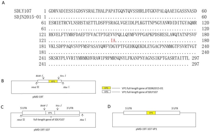
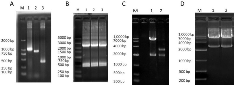
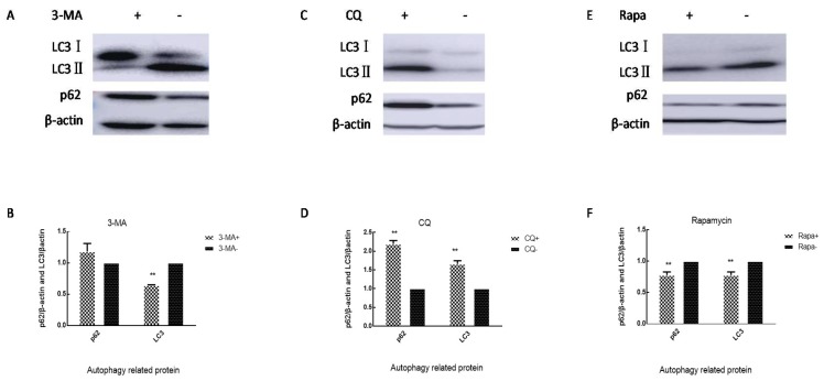
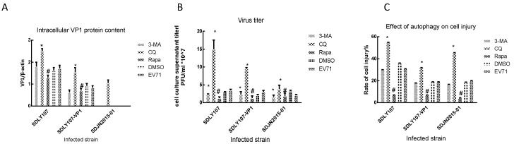
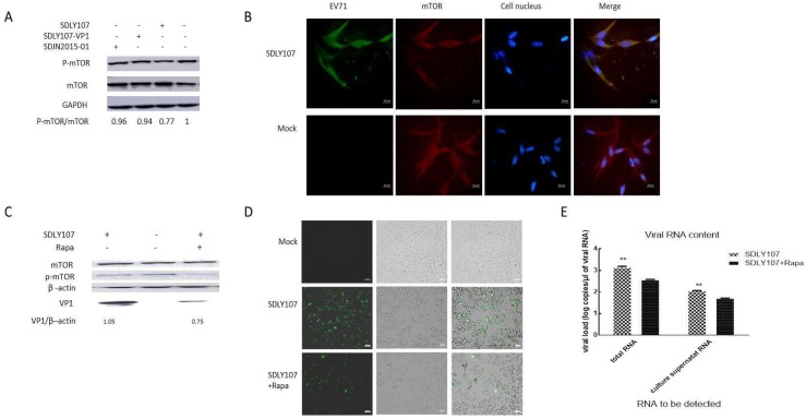
Methods: A recombinant virus-SDLY107-VP1, obtained by replacing the VP1 full-length gene of the SDLY107 strain with the VP1 full-length gene of the attenuated strain SDJN2015-01-was constructed and tested for replication and virulence. We then tested the effect of the recombinant virus on autophagy in nerve cells. The effect of autophagy on virus replication was detected by western blot and plaque test. Finally, the changes of mTOR signaling molecules during EV71 infection and the effect of mTOR on virus replication at the RNA level were detected.

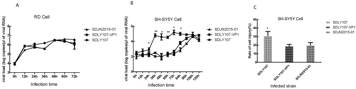
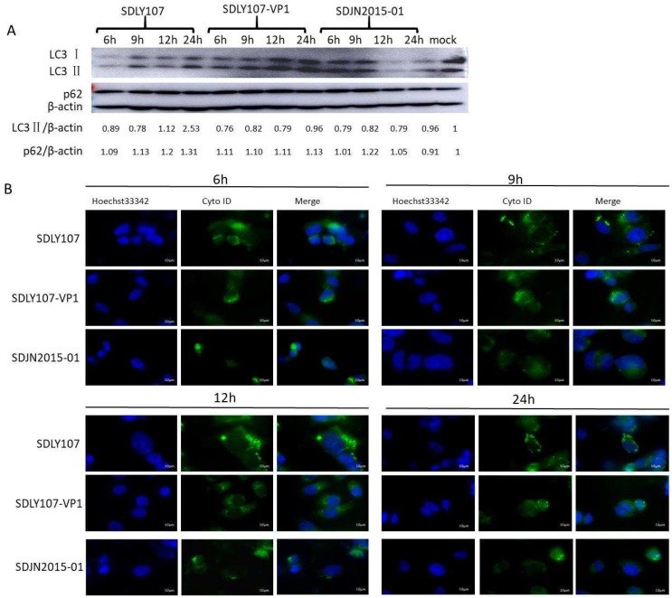
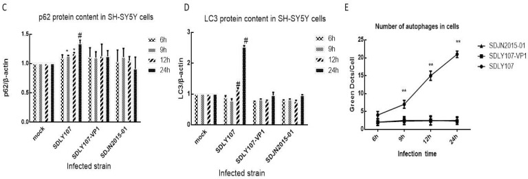
Results: Viral recombination triggered virulence attenuation. The replication ability of recombinant virus SDLY107-VP1 was significantly weaker than that of the parent strain SDLY107. The SDLY107 strain could inhibit autophagic flux and led to accumulation of autophagosomes, while the SDLY107-VP1 strain could not cause autophagosome accumulation. The synthesis of EV71 RNA was inhibited by inhibiting mTOR.

Conclusions: Replacement of VP1 weakened the replication ability of virulent strains and reduced the level of autophagy in nerve cells. This autophagy facilitates the replication of virulent strains in nerve cells. VP1 is an important neurovirulence determinant of EV71, which affects virus replication by regulating cell autophagy. mTOR is a key molecule in this type of autophagy.

Keywords: EV71; VP1; autophagy; virus replication.


Conflict of interest statement

The authors declare no conflict of interest. The funders had no role in the design of the study in the collection, analyses, or interpretation of data, in the writing of the manuscript, or in the decision to publish the results.

Figures

Figure 1
Figure 1
Schematic diagram of SDLY107-VP1. (A) The VP1 amino acid sequence comparison results of SDLY107 and SDJN2015-01 strains are shown. (B) The upstream and downstream fragments of VP1 of SDLY107 strain and VP1 of SDJN2015-01 strain were used as templates for fusion PCR. The obtained recombinant DNA fragments were linked to the pMD-19T vector and identified by double digestion. (C) The full-length pMD-19T-107 plasmid was identified by double digestion, it contains a full-length gene including the untranslated region (UTR) of SDLY107 strain. (D) DNA fragments containing the VP1 sequence of the SDJN2015-01 strain and the remaining DNA fragments without the VP1 fragment of the SDLY107 strain were recovered, then transformed after T4 connection, and the recombinant plasmid pMD-19T-107-VP1 was successfully obtained.
Figure 2
Figure 2
Recombinant virus SDLY107-VP1. (A) The PCR product of the 1430–2459 bp upstream fragment of VP1 of the SDLY107 strain, the 3308–3742 bp downstream fragment, and the VP1 fragment of the SDJN2015-01 strain. M is DL2000 DNA Marker; Lane 1 is upstream 1430–2459 bp of VP1 of the SDLY107 strain, and the length is about 1029 bp; Lane 2 is VP1 fragment of the SDJN2015-01 strain, and the length is about 891 bp; Lane 3 is downstream 3308–3742 bp of VP1 of the SDLY107 strain, and the length is approximately 434 bp. (B) The fusion products of the 1430–2459 bp upstream fragment, 3308–3742 bp downstream fragment and VP1 fragment of the SDJN2015-01 strain were obtained. M is a DL5000 DNA Marker; Lanes 1–3 are three DNA fragment fusion PCR products, and the length is about 2313 bp. (C) Plasmid pMD-19T-107 and pMD-19T-VP1 double digestion enzyme electrophoresis. M is DL1,0000 DNA Marker; Lane 1 is the product of the pMD-19T-107 plasmid after Nru I and BsiW I digestion, the fragment length is about 8.1 kb and 2.0 kb; Lane 2 is the pMD-19T-VP1 plasmid after Nru I and BsiW I digestion. The product fragment length is approximately 3.0 kb and 2.0 kb. (D) Plasmid pMD-19T-107-VP1 double digestion enzyme electrophoresis. M is DL1,0000 DNA Marker; Lanes 1 and 2 are the product of the pMD-19T-107-VP1 plasmid digested with XbaI and HindIII, and the fragment length is about 7.4 kb and 2.7 kb.
Figure 3
Figure 3
(A)The replication curves of the three strains on rhabdomyosarcoma (RD) cells. (B) The replication curves of the three strains on SH-SY5Y cells. (C)The rate of cell injury of SH-SY5Y cells infected with the three strains at 24 h. The data represent means + SD from three experiments. * p < 0.05, ** p < 0.001 compared with the SDLY107-VP1 treated group.
Figure 4
Figure 4
(A) Western blot assay for the effect of recombinant virus SDLY107-VP1 strain, parental virus SDLY107 strain, and SDJN2015-01 strain on autophagy. SH-SY5Y cells were infected with EV71. Cell lysates were collected at 6 h, 9 h, 12 h, and 24 h. Uninfected cells served as the sham control. LC3 and p62 were examined by western blot. (B) The effects of the recombinant virus SDLY107-VP1 strain, parental virus SDLY107 strain, and SDJN2015-01 strain on autophagy were detected by immunofluorescent staining. Hoechst 33342 blue fluorescent dye stains the cell nucleus blue, and Cyto ID green fluorescent dye stains autophagous bodies green during autophagy. (C) The content of p62 protein in SH-SY5Y cells infected with the three viruses. (D) The content of LC3 protein in SH-SY5Y cells infected with the three viruses. (E) The number of autophages in SH-SY5Y cells infected with the three viruses. Autophagous bodies in each cell were counted, 100 cells per sample. The data represent means + SD from three experiments. * p < 0.05, compared with the mock group; # p < 0.001, compared with the mock group; ** p < 0.05, compared with the SDLY107-VP1-treated group.
Figure 4
Figure 4
(A) Western blot assay for the effect of recombinant virus SDLY107-VP1 strain, parental virus SDLY107 strain, and SDJN2015-01 strain on autophagy. SH-SY5Y cells were infected with EV71. Cell lysates were collected at 6 h, 9 h, 12 h, and 24 h. Uninfected cells served as the sham control. LC3 and p62 were examined by western blot. (B) The effects of the recombinant virus SDLY107-VP1 strain, parental virus SDLY107 strain, and SDJN2015-01 strain on autophagy were detected by immunofluorescent staining. Hoechst 33342 blue fluorescent dye stains the cell nucleus blue, and Cyto ID green fluorescent dye stains autophagous bodies green during autophagy. (C) The content of p62 protein in SH-SY5Y cells infected with the three viruses. (D) The content of LC3 protein in SH-SY5Y cells infected with the three viruses. (E) The number of autophages in SH-SY5Y cells infected with the three viruses. Autophagous bodies in each cell were counted, 100 cells per sample. The data represent means + SD from three experiments. * p < 0.05, compared with the mock group; # p < 0.001, compared with the mock group; ** p < 0.05, compared with the SDLY107-VP1-treated group.
Figure 5
Figure 5
Cells were treated with 3-MA 50 μM, CQ 20 μM, and rapamycin 10 nM. After 2 h, cells were washed three times, and the levels of p62 and LC3 in the cells were measured after 24 h. (A) Western blot detects the effect of 3-MA on autophagy in SH-SY5Y cells. (B) Changes in intracellular p62 and LC3 levels in SH-SY5Y cells after 3-MA treatment of cells. (C) Western blot detects the effect of CQ on autophagy in SH-SY5Y cells. (D) Changes in intracellular p62 and LC3 levels in SH-SY5Y cells after CQ treatment of cells. (E) Western blot detects the effect of rapamycin on autophagy in SH-SY5Y cells. (F) Changes in intracellular p62 and LC3 levels in SH-SY5Y cells after rapamycin treatment of cells. The data represent means + SD from three experiments, ** p < 0.001, compared with the control group.
Figure 6
Figure 6
(A) Intracellular VP1 protein content. SH-SY5Y cells were treated with 3-MA, CQ, or rapamycin for 2 h and then infected with SDLY107, SDLY107-VP1, and SDJN2015-01, respectively. Cell lysates were harvested after 24 h. LC3, p62, and capsid protein VP1 levels were detected by western blot. (B) Virus titer of culture supernatant. SH-SY5Y cells were treated with 3-MA, CQ, or rapamycin for 2 h and then infected with SDLY107, SDLY107-VP1, and SDJN2015-01, respectively. After 24 h, the cell culture supernatant was harvested and the culture supernatant titer was measured by plaque assay. (C) Effect of autophagy on cell injury. SH-SY5Y cells were treated with 3-MA, CQ, or rapamycin for 2 h and then infected with SDLY107, SDLY107-VP1, and SDJN2015-01, respectively. After 24 h, LDH activity was assayed using the Cytotoxicity Assay Kit according to the manufacturer’s protocol. Rapa stands for rapamycin. The data represent means +SD from three experiments. * p < 0.05, compared with the EV71-treated group; # p < 0.05, compared with the DMSO-treated group.
Figure 7
Figure 7
(A) SH-SY5Y cells were infected with the three strains. Cell lysates were collected at 24 h. mTOR and p-mTOR levels were detected by western blot. (B) EV71 and mTOR were detected by co-localization immunofluorescence at 24 h after infection. FICT stained EV71 green, TRITC stained mTOR red, and DAPI stained nucleus blue. (C) SH-SY5Y cells were seeded on a six well plate and exposed to rapamycin or not before being infected with EV71. The viral VP1 protein was examined by western blot after 24 h. (D) SH-SY5Y cells were inoculated into a 12 well plate with about 2 × 10⁵ cells per well and exposed to rapamycin or not before being infected with EV71. Immunofluorescent assay was used to detect the intracellular virus content after 24 h. (E) SH-SY5Y cells were inoculated onto a 12 well plate and exposed to rapamycin or not before being infected with EV71, and the amount of viral RNA was measured by real-time fluorescent quantitative PCR after 24 h. Rapa stands for rapamycin. The data represent means + SD from three experiments, ** p < 0.001, compared with the rapamycin treatment group.

Similar articles

Cited by

References

    1. Ayukekbong J.A., Bergstrom T. Polio will go, acute flaccid paralysis will stay. Lancet. 2014;383:2209–2210. doi: 10.1016/S0140-6736(14)61080-1. - DOI - PubMed
    1. Nassef C., Ziemer C., Morrell D.S. Hand-foot-and-mouth disease: A new look at a classic viral rash. Curr. Opin. Pediatr. 2015;27:486–491. doi: 10.1097/MOP.0000000000000246. - DOI - PubMed
    1. Wu Y., Yeo A., Phoon M.C., Tan E.L., Poh C.L., Quak S.H., Chow V.T. The largest outbreak of hand; foot and mouth disease in Singapore in 2008: The role of enterovirus 71 and coxsackievirus A strains. Int. J. Infect. Dis. 2010;14:1076–1081. doi: 10.1016/j.ijid.2010.07.006. - DOI - PubMed
    1. Chan L.G., Parashar U.D., Lye M.S., Ong F.G., Zaki S.R., Alexander J.P., Ho K.K., Han L.L., Pallansch M.A., Suleiman A.B., et al. Deaths of children during an outbreak of hand, foot, and mouth disease in sarawak, malaysia: Clinical and pathological characteristics of the disease. For the Outbreak Study Group. Clin. Infect. Dis. 2000;31:678–683. doi: 10.1086/314032. - DOI - PubMed
    1. Van Tu P., Thao N.T.T., Perera D., Truong K.H., Tien N.T.K., Thuong T.C., How O.M., Cardosa M.J., McMinn P.C. Epidemiologic and virologic investigation of hand, foot, and mouth disease, southern Vietnam, 2005. Emerg. Infect. Dis. 2007;13:1733–1741. doi: 10.3201/eid1311.070632. - DOI - PMC - PubMed

Publication types

LinkOut - more resources