

Anti-Wrinkle Benefits of Peptides Complex Stimulating Skin Basement Membrane Proteins Expression
- PMID: 31861912
- PMCID: PMC6981886
- DOI: 10.3390/ijms21010073
Anti-Wrinkle Benefits of Peptides Complex Stimulating Skin Basement Membrane Proteins Expression
Abstract
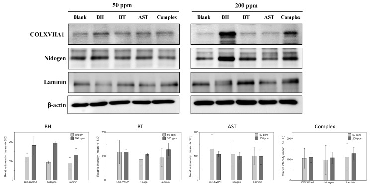
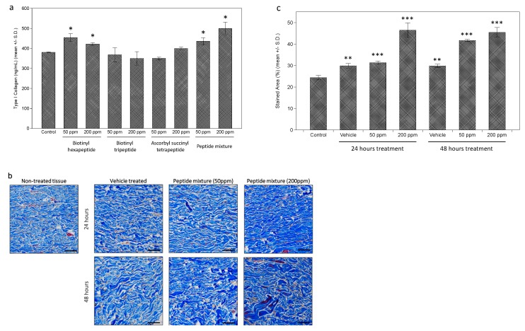
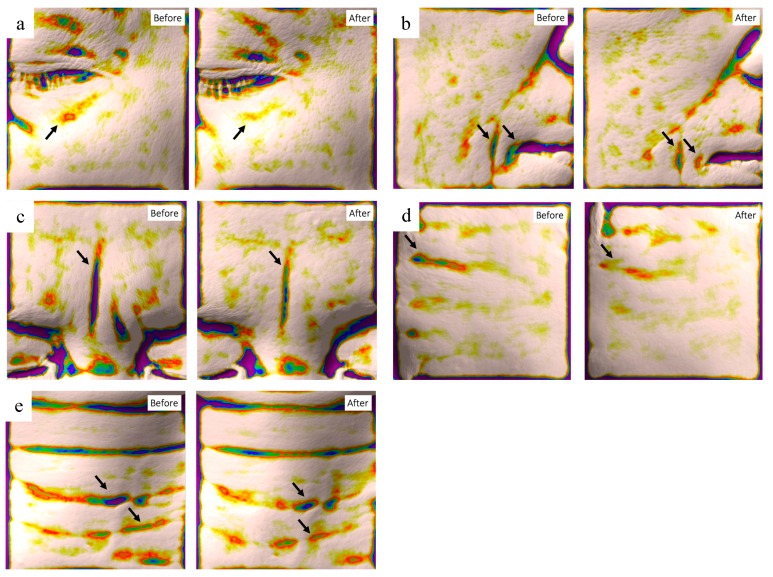
The dermal-epidermal junction (DEJ) provides a physical and biological interface between the epidermis and the dermis. In addition to providing a structural integrity, the DEJ also acts as a passageway for molecular transport. Based on the recently reported importance of the DEJ in skin aging, novel peptide derivatives have been tested for their effects on basement membrane (BM) protein expressions in cultured human epidermal keratinocytes. As a result, protein expressions of collagen XVII, laminin and nidogen were stimulated by the test peptide and peptides complex. Further ex vivo evaluation using excised human skin, confirmed that the topical application of the peptides complex significantly increased dermal collagen expression, as well as expressions of collagen XVII and laminin. Interestingly, while the origin of the laminin protein is epidermal keratinocytes, the immunohistochemical staining of skin showed that laminin was only detected in the uppermost layer of the dermis, which suggests a tight assembly of laminin protein onto the dermal side of the DEJ. These results suggest that a peptide complex could improve the structural properties of the DEJ through its ability to stimulate BM proteins. In order to evaluate the anti-wrinkle benefits of the peptide complex in vivo, a clinical study was performed on 22 healthy Asian female volunteers older than 40 years. As a result, significant improvements in skin wrinkles for all of the five sites were observed after two weeks, as assessed by skin topographic measurements. Collectively, these results demonstrate the anti-aging efficacy of the peptides complex.
Keywords: clinical efficacy; dermal-epidermal junction; laminin; nidogen; peptides complex; wrinkle.
Conflict of interest statement
Incospharm Corp. and Chameditech Corp. design and manufacture test peptides (biotinyl hexapeptide and biotinyl tripeptide from Incospharm Corp and Ascorbyl succinyl tetrapeptide from Chameditech Corp). P&K Skin Research Center Co., Ltd., performed clinical efficacy testing. Cha BIO F&C made a tested formulation with peptide complex and currently sells cosmetic products containing tested peptide complex.
Figures

Similar articles
-
Synergistic action of a triple peptide complex on an essential extra-cellular matrix protein exhibits significant anti-aging benefits.J Cosmet Dermatol. 2010 Jun;9(2):108-16. doi: 10.1111/j.1473-2165.2010.00494.x. J Cosmet Dermatol. 2010. PMID: 20618556 Clinical Trial.
-
Improvement of the dermal epidermal junction in human reconstructed skin by a new c-xylopyranoside derivative.Eur J Dermatol. 2008 May-Jun;18(3):297-302. doi: 10.1684/ejd.2008.0392. Epub 2008 May 13. Eur J Dermatol. 2008. PMID: 18474459
-
The effects of epidermal keratinocytes and dermal fibroblasts on the formation of cutaneous basement membrane in three-dimensional culture systems.Arch Dermatol Res. 2005 Jan;296(7):296-302. doi: 10.1007/s00403-004-0529-5. Epub 2004 Dec 10. Arch Dermatol Res. 2005. PMID: 15650892
-
Importance of the dermal-epidermal junction and recent advances.Dermatologica. 1987;174(1):1-10. doi: 10.1159/000248972. Dermatologica. 1987. PMID: 3542613 Review.
-
Epidermal basement membrane zone components: ultrastructural distribution and molecular interactions.J Dermatol Sci. 2003 May;31(3):169-77. doi: 10.1016/s0923-1811(03)00045-8. J Dermatol Sci. 2003. PMID: 12727020 Review.
Cited by
-
Wrinkle-Improving Effect of Novel Peptide That Binds to Nicotinic Acetylcholine Receptor.Int J Mol Sci. 2024 Jul 18;25(14):7860. doi: 10.3390/ijms25147860. Int J Mol Sci. 2024. PMID: 39063099 Free PMC article.
-
Cosmeceutical Peptides in the Framework of Sustainable Wellness Economy.Front Chem. 2020 Oct 30;8:572923. doi: 10.3389/fchem.2020.572923. eCollection 2020. Front Chem. 2020. PMID: 33195061 Free PMC article. Review.
-
Physicochemical and Antioxidative Properties of Protein Hydrolysates from Residual Goat Placenta Extract by Two Different Methods.Foods. 2024 Oct 14;13(20):3263. doi: 10.3390/foods13203263. Foods. 2024. PMID: 39456325 Free PMC article.
-
Design and Evaluation of Complex Polypeptide-Loaded Dissolving Microneedles for Improving Facial Wrinkles in Different Areas.Polymers (Basel). 2022 Oct 22;14(21):4475. doi: 10.3390/polym14214475. Polymers (Basel). 2022. PMID: 36365468 Free PMC article.
-
Ectoin attenuates cortisone-induced skin issues by suppression GR signaling and the UVB-induced overexpression of 11β-HSD1.J Cosmet Dermatol. 2024 Dec;23(12):4303-4314. doi: 10.1111/jocd.16516. Epub 2024 Sep 2. J Cosmet Dermatol. 2024. PMID: 39222375 Free PMC article.
References
-
- Newton V.L., Bradley R.S., Seroul P., Cherel M., Griffiths C.E.M., Rawlings A.V., Voegeli R., Watson R.E.B., Sherratt M.J. Novel approaches to characterize age-related remodelling of the dermal-epidermal junction in 2D, 3D and in vivo. Skin Res. Technol. 2017;23:131–148. doi: 10.1111/srt.12312. - DOI - PubMed
MeSH terms
Substances
LinkOut - more resources
Full Text Sources
Other Literature Sources
Medical
Research Materials
