. 2020 Feb 5;28(2):631-641.
doi: 10.1016/j.ymthe.2019.11.025. Epub 2019 Dec 6.

Upregulation of PUM1 Expression in Preeclampsia Impairs Trophoblast Invasion by Negatively Regulating the Expression of the lncRNA HOTAIR

Affiliations

Upregulation of PUM1 Expression in Preeclampsia Impairs Trophoblast Invasion by Negatively Regulating the Expression of the lncRNA HOTAIR

Yan Zhang et al. Mol Ther. .

Abstract

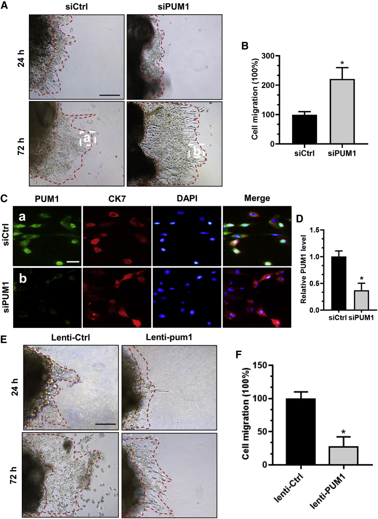
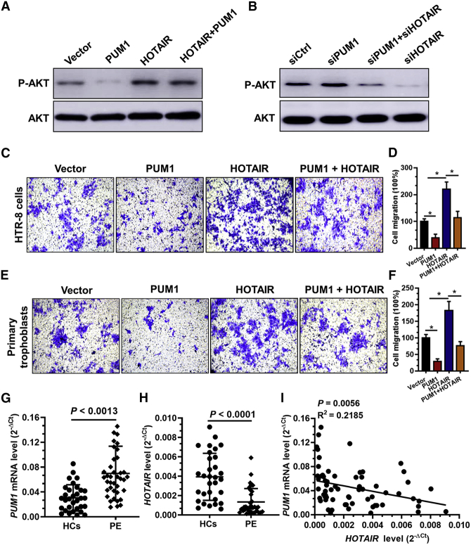
Pumilio (PUM) proteins are members of a highly conserved RNA-binding protein family that posttranscriptionally regulate gene expression in many organisms. However, their roles in the placenta are unclear. In the present study, we report the requirement for the PUM homolog 1 (PUM1) gene in preeclampsia (PE). Immunofluorescence and immunohistochemical data showed that PUM1 was highly expressed in human placental villi from women with PE compared to healthy controls (HCs). Further, PUM1 overexpression repressed, and knockdown enhanced, the invasion and proliferation of trophoblasts. Interestingly, PUM1 knockdown promoted trophoblast invasion in a villous explant culture model, while PUM1 overexpression repressed these effects. Furthermore, lncRNA transcriptome sequencing coupled with RNA immunoprecipitation (RIP) revealed that PUM1 inhibits trophoblast invasion in PE by downregulating the expression of lncRNA HOTAIR. Moreover, PUM1 regulates HOTAIR expression via a posttranscriptional mechanism. Using RNA-protein pull-down and mRNA stability assays, we identified PUM1 as a specific binding partner that decreased the half-life of HOTAIR and lowered the steady-state level of HOTAIR expression, suggesting a novel posttranscriptional regulatory mechanism. Collectively, these findings identified a novel RNA regulatory mechanism, revealing a new pathway governing the regulation of PUM1/HOTAIR in trophoblast invasion in the pathogenesis of PE.

Keywords: HOTAIR; PUM1; lncRNA stability; preeclampsia; trophoblast invasion.


Figures

Figure 1
Figure 1
PUM1 Is Upregulated in Placental Trophoblasts Isolated from Preeclampsia (PE) Patients (A) PUM1 expression in human placental villi tissues from PE patients (n = 12) and in healthy controls (HCs) (n = 12) was determined by qRT-PCR. (B) Level of PUM1 expression in human placental villi tissues from PE patients and in HCs was determined by western blotting analysis. (C) Single staining of maternal villi from HCs (n = 29), early-onset PE (n = 10), and late-onset PE patients (n = 27) was performed using rabbit IgG anti-human-PUM1 antibody and developed with a labeled streptavidin biotin+ horseradish peroxidase (HRP) kit. The sections were counterstained with hematoxylin, and positive cells were quantified using ImagePro-plus 6.0 software (D). *p < 0.05 compared with HCs. Scale bars, 100 μm. (E) Representative immunofluorescence of PUM1 in primary trophoblasts from villi tissue of late-onset PE patients (n = 6) and HCs (n = 6). Fluorescence signals specific to anti-PUM1 antibodies appear green, and the DAPI-stained nuclei appear blue. Scale bar, 25 μm. (F) Fluorescence intensity of the signaling was assessed by Leica confocal SP8 software. *p < 0.05 compared with HCs.
Figure 2
Figure 2
PUM1 Decreases Trophoblast Proliferation and Invasion (A) Western blotting analysis of PUM1 expression in HTR-8 cells transfected with short interfering RNAs siCtrl, siPUM1 #1, siPUM1 #2, siPUM1 #3, control vector, or PUM1-expressing vector after 48 h. (B) HTR-8 cells were transfected with one of the above-mentioned siPUM1 #3 or vectors and incubated for 48 h. Cell proliferation was measured using the cell counting kit-8 (CCK-8) assay at the indicated times. *p < 0.05 compared with siCtrl or vector. (C) PUM1 knockdown drastically promoted the rate of wound closure compared with that of the scrambled control cell line. Overexpression of PUM1 in HTR-8 cells resulted in decreased wound closure ability compared with that in the vector control cells. Original magnification ×100. (D and E) Overexpression of PUM1 in HTR-8 cells significantly reduced cell invasion compared with that of the vector control cell line (D). The invasive ability of the cells was assessed by crystal violet staining (E); original magnification ×200. *p < 0.05 compared with the control vector. (F and G) Knockdown of PUM1 increased cell invasion compared with that of the scrambled control cell line (F). The invasive ability of the cells was assessed by crystal violet staining (G); original magnification ×200. *p < 0.05 compared with the siCtrl.
Figure 3
Figure 3
PUM1 Reduced Trophoblast Outgrowth in Extravillous Explant Cultures (A) Extravillous explants were maintained in culture on Matrigel. Serial pictures of the explants incubated with siCtrl or siPUM1 #3 oligos were taken under a light microscope after 24 h and 72 h of culture in vitro. Scale bar, 250 μm. (B) Statistical assay of the migration distance of villous tips (%). *p < 0.05 compared with the siCtrl. (C) The extravillous explants were cultured on Matrigel for 72 h. Immunofluorescence staining using anti-PUM1 antibodies showed an obvious decrease in the PUM1 protein level in the short interfering RNA siPUM1 group compared with the siCtrl group. Green fluorescence signals indicate bound anti-PUM1 antibodies; CK7 staining is visualized as red; and a DAPI-stained nucleus is blue. Scale bar, 25 μm. (D) The level of PUM1 expression was assessed using Leica confocal SP8 software. *p < 0.05 compared with the siCtrl. (E) Extravillous explants from healthy controls (HCs) were maintained in culture on Matrigel. Serial pictures of the explants incubated with lenti-ctrl or lenti-PUM1 lentivirus were taken under a light microscope after 24 and 72 h of culture in vitro. Scale bar, 250 μm. (F) Statistical assay of the migration distance of villous tips (%). *p < 0.05 compared with the lenti-ctrl.
Figure 4
Figure 4
PUM1 Reduced HOTAIR Expression in Trophoblasts (A) Heatmap of the normalized expression levels of lncRNAs in HTR-8 cells transfected with short interfering RNAs siCtrl or siPUM1-3. Blue indicates downregulation expression levels; red indicates upregulation expression levels. (B) lncRNAs sequencing results showing the expression levels of significantly different genes. siPUM1 compared to siCtrl. (C) Real-time PCR was performed to determine the levels of the typical lncRNAs in HTR-8 cells transfected with siCtrl or siPUM1. *p < 0.05 compared with the siCtrl. (D) Real-time PCR was performed to determine the mRNA levels of the typical lncRNAs in HTR-8 cells transfected with the vector or the PUM1 overexpression vector. *p < 0.05 compared with the vector. (E) Analysis of co-purified RNA and respective enrichment as determined by real-time qRT-PCR after anti-PUM1 immunoprecipitation to validate the specific binding of the indicated lncRNAs to PUM1 in HTR-8 cells. All immunoprecipitation experiments were performed using three biological replicates. *p < 0.05 compared with input. (F and G) qRT-PCR analysis of HOTAIR expression in HTR-8 cells transfected with siRNAs siCtrl, siPUM1 #2, siPUM1 #3 (F), control vector, or PUM1 overexpression vector (G) after 48 h. *p < 0.05 compared with the vector or siCtrl.
Figure 5
Figure 5
PUM1 Is an Important RNA Binding Protein of HOTAIR in Trophoblasts (A) The diagram illustrates the PUM1 binding site in the primary transcript region of HOTAIR on the human chromosome. The human DNA sequences of putative PUM1 binding sites are labeled as ARE 1–4, and the core element (TGTARATA) for PUM1 binding is indicated in red. The mutant PUM1 binding sites are labeled as MUT 1–4, and the mutant element (AACTRATA) for PUM1 binding is also indicated in red. (B) A schema for the constructs and co-transfection experiments. (C) A fragment of the sixth exon region of HOTAIR was cloned into a psiCheck2 vector, downstream of the renilla luciferase gene. These constructs where co-transfected with the siPUM1 or siCtrl siRNA into HTR-8 cells. The cells were harvested after 24 h. Renilla luciferase activity was measured and normalized to that of firefly luciferase. The results are represented as the mean of three independent experiments. *p < 0.05 compared with siCtrl. (D) HTR-8 cells were transfected with 35-bp HOTAIR antisense oligos (A–D and C+D), which contained a core element (UGUARAUA) for PUM1 binding, and 35-bp unrelated in-sequence oligos for 12 h. Analysis of co-purified RNA and respective enrichment as detected by qRT-PCR after anti-PUM1 immunoprecipitation to validate the enrichment of HOTAIR to PUM1 in HTR-8 cells. All immunoprecipitation experiments were performed using three biological replicates. (E) Validation of specific binding of PUM1 to biotinylated HOTAIR in HTR-8 cell by western blotting. (F and G) HOTAIR stability analysis in HTR-8 cells after actinomycin D (5 μg/mL) treatment. Cells were transfected with the PUM1 overexpression plasmid (F) or siPUM1 (G) for 36 h; a time course for RNA stability was initiated by adding the RNA-polymerase II inhibitor (actinomycin D ActD]). The cells were harvested at the indicated time points. Expression levels were normalized to “0 h,” and glyceraldehyde-3-phosphate dehydrogenase (GAPDH mRNA) was used as the reference gene. The results are shown as the mean of at least three independent experiments. *p < 0.05 compared with the vector or siCtrl.
Figure 6
Figure 6
The PUM1/HOTAIR/AKT Pathway Is Involved in Regulation of Trophoblast Invasion (A) HTR-8 cells were transfected with control vector or PUM1-, HOTAIR-, or PUM1+HOTAIR-expressing vectors after 48 h. The expression levels of phosphorylated (P)-AKT ser473 and AKT were determined using western blotting. (B) HTR-8 cells were transfected with short interfering RNAs siCtrl, siHOTAIR, siPUM1, or siHOTAIR+siPUM1 after 48 h. The levels of p-AKT ser473 and AKT expression were determined using western blotting. (C–F) HTR-8 cells (C) and primary trophoblasts (E) from first trimester villous were transfected with control vector or PUM1-, HOTAIR-, PUM1+HOTAIR-expressing vectors after 48 h. The invasive ability of HTR-8 cells (D) and primary trophoblasts (F) was assessed using ImagePro-plus 6.0., (G and H) Level of PUM1 mRNA (G) and HOTAIR (H) expression in human placental villi tissues from preeclampsia (PE) (n = 41) patients and in healthy controls (HCs; n = 39) were determined by qRT-PCR. (I) The HOTAIR level in the villous tissue of PE patients was measured using qRT-PCR and correlated with the PUM1 mRNA level in the villous tissue of the corresponding patients.

Similar articles

Cited by

References

    1. Sharma S., Godbole G., Modi D. Decidual Control of Trophoblast Invasion. Am. J. Reprod. Immunol. 2016;75:341–350. - PubMed
    1. Aplin J.D. Implantation, trophoblast differentiation and haemochorial placentation: mechanistic evidence in vivo and in vitro. J. Cell Sci. 1991;99:681–692. - PubMed
    1. Staun-Ram E., Shalev E. Human trophoblast function during the implantation process. Reprod. Biol. Endocrinol. 2005;3:56–63. - PMC - PubMed
    1. Strickland S., Richards W.G. Invasion of the trophoblasts. Cell. 1992;71:355–357. - PubMed
    1. Lain K.Y., Roberts J.M. Contemporary concepts of the pathogenesis and management of preeclampsia. JAMA. 2002;287:3183–3186. - PubMed

Publication types