. 2019 Dec 20;10(12):956.
doi: 10.1038/s41419-019-2181-1.

Paclitaxel induces lymphatic endothelial cells autophagy to promote metastasis

Affiliations

Paclitaxel induces lymphatic endothelial cells autophagy to promote metastasis

Audrey Zamora et al. Cell Death Dis. .

Abstract

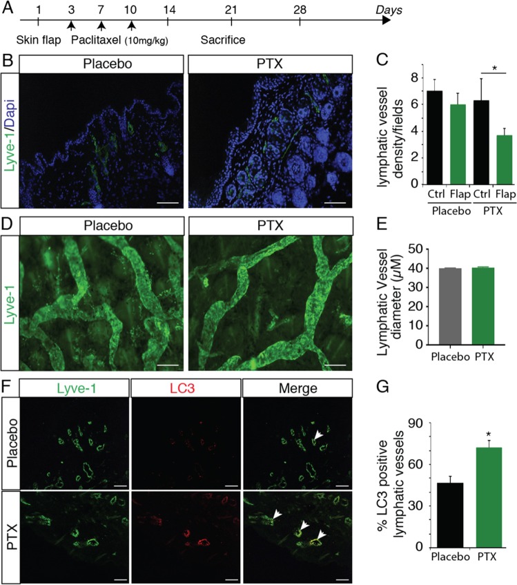
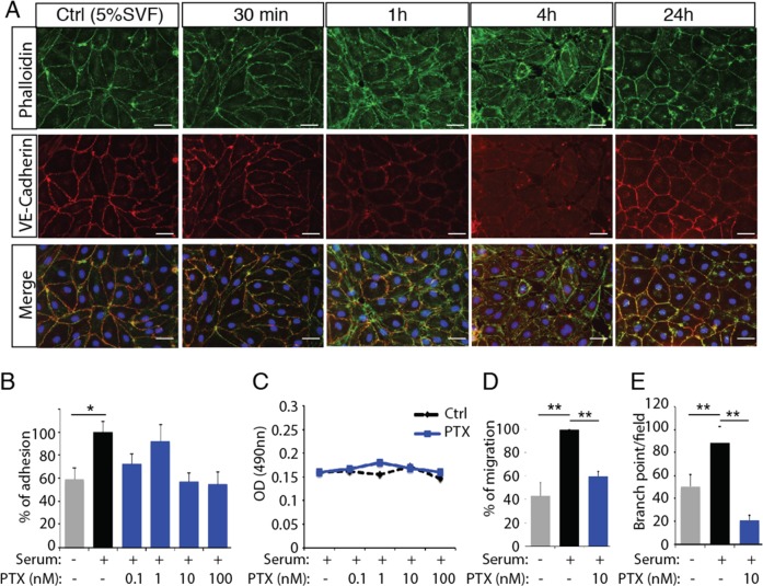
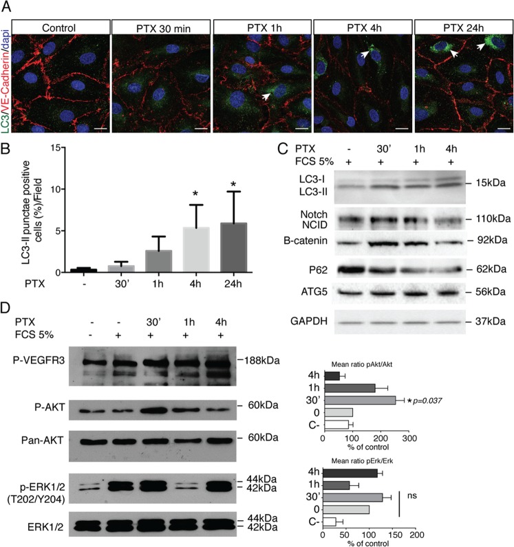
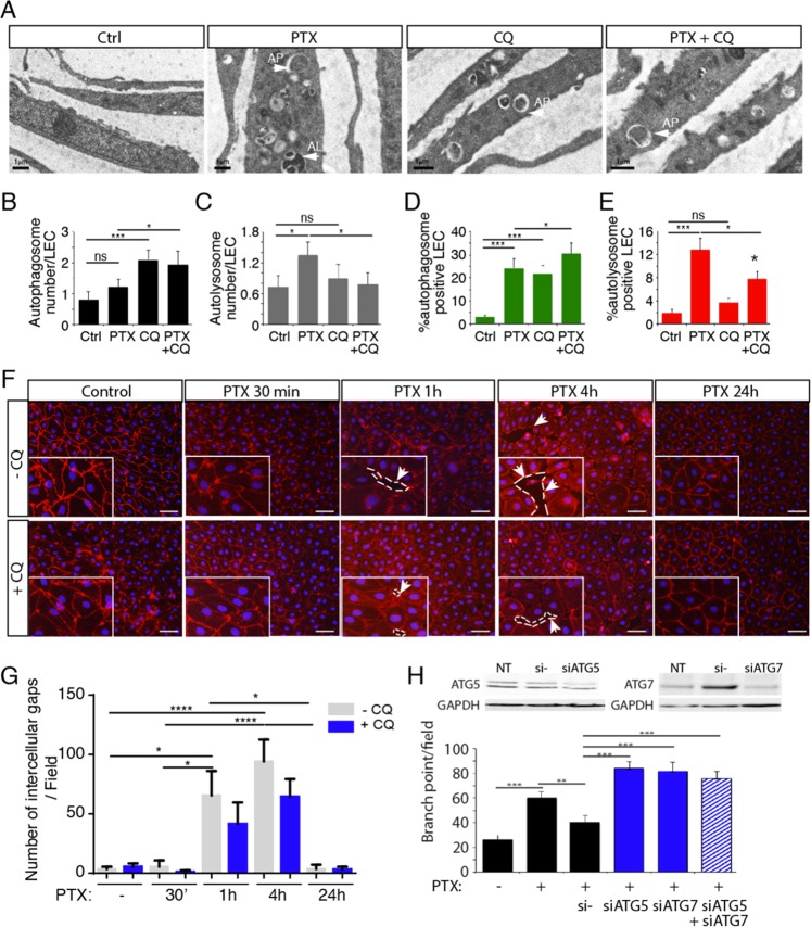
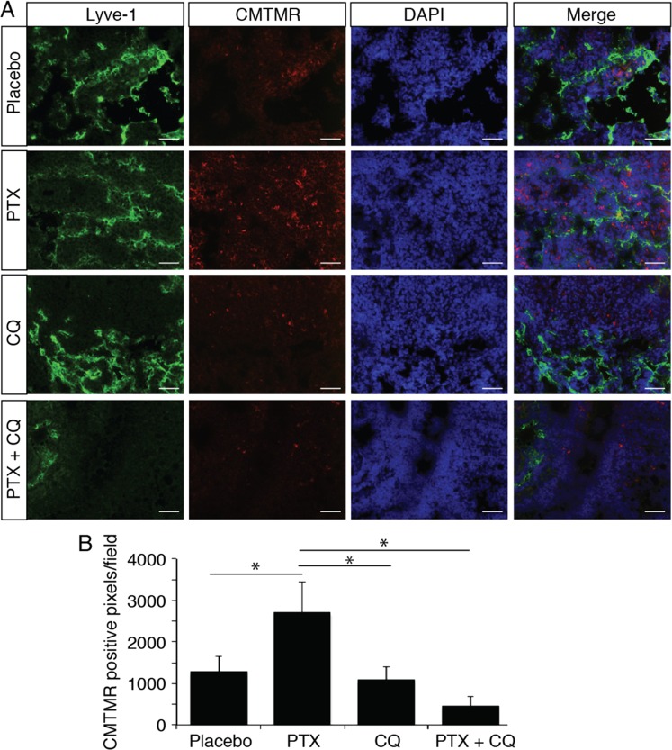
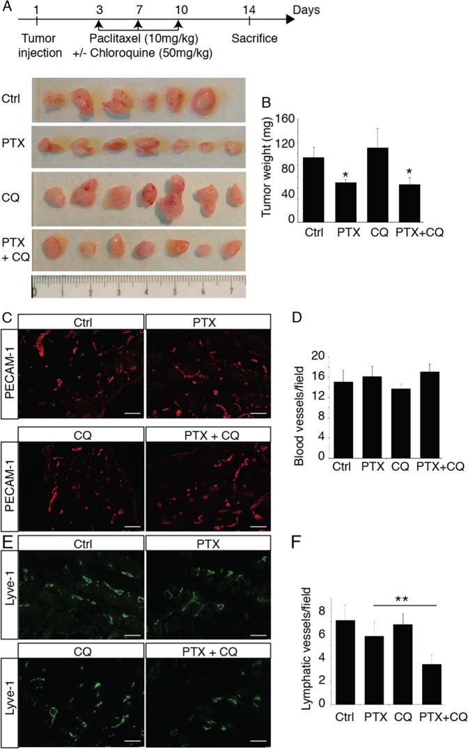
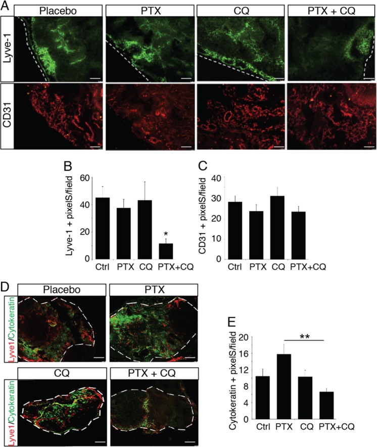
Cytotoxic therapy for breast cancer inhibits the growth of primary tumors, but promotes metastasis to the sentinel lymph nodes through the lymphatic system. However, the effect of first-line chemotherapy on the lymphatic endothelium has been poorly investigated. In this study, we determined that paclitaxel, the anti-cancer drug approved for the treatment of metastatic or locally advanced breast cancer, induces lymphatic endothelial cell (LEC) autophagy to increase metastases. While paclitaxel treatment was largely efficacious in inhibiting LEC adhesion, it had no effect on cell survival. Paclitaxel inhibited LEC migration and branch point formation by inducing an autophagy mechanism independent of Akt phosphorylation. In vivo, paclitaxel mediated a higher permeability of lymphatic endothelium to tumor cells and this effect was reversed by chloroquine, an autophagy-lysosome inhibitor. Despite a strong effect on reducing tumor size, paclitaxel significantly increased metastasis to the sentinel lymph nodes. This effect was restricted to a lymphatic dissemination, as chemotherapy did not affect the blood endothelium. Taken together, our findings suggest that the lymphatic system resists to chemotherapy through an autophagy mechanism to promote malignant progression and metastatic lesions. This study paves the way for new combinative therapies aimed at reducing the number of metastases.


Conflict of interest statement

The authors declare that they have no conflict of interest.

Figures

Fig. 1
Fig. 1. PTX reduces lymphatic healing.
a Schematic representation of the skin flap experimental procedure. b Representative fluorescent images of Lyve-1-positive regenerated lymphatic vessels from PTX-treated mice 4 weeks after skin flap. Scale bar: 50 μm. c Quantification of the lymphatic vessel density in PTX-treated mice, *p < 0.05. d Immunostaining for Lyve-1 shows no difference in lymphatic vessel size in PTX-treated mice. Scale bar: 200 μm. e Quantification of the lymphatic vessel diameter in PTX-treated mice skin. f Immunostaining for Lyve-1 (green) and LC3 (red) shows increase of LC3 expression in lymphatic vessels from PTX-treated mice. Scale bar: 50 μm. g Quantification of the percentage of LC3-positive lymphatic vessels in PTX-treated mice skin.
Fig. 2
Fig. 2. PTX inhibits lymphatic endothelial cell function in vitro.
a Phalloidin (green) and VE-cadherin (red) immunostaining on HDLEC stimulated by PTX (10 nM) for 30 min, 1 h, 4 h, and 24 h shows a downregulation of VE-cadherin expression after 1 h and 4 h associated with an alteration of cortical actin organization at cellular junctions as show with phalloidin immunostaining. Scale bar 50 μm. b, c Increasing doses of PTX (0.1–100 nM) reduced HDLEC adhesion in the presence of 5% SVF without significant effect on HDLEC survival as shown by MTT assay. d Quantification of the percentage of HDLEC migration using scratch wound healing assay shows an inhibition of serum-induced (5%SVF) migration by PTX. **p < 0.01. e PTX antagonizes serum-induced (5%SVF) HDLEC branching in matrigel. **p < 0.01.
Fig. 3
Fig. 3. PTX induces HDLEC autophagy.
a LC3 (green) and VE-cadherin (red) immunostaining on HDLEC stimulated by PTX (10 μM) for 30 min, 1 h, 4 h, and 24 h shows an upregualtion of LC3 dots after 1 h, 4 h and 24 h of stimulation. b Quantification of the LC3-positive HDLEC treated with PTX. *p < 0.05. c Immunoblot analysis of LC3-I, LC3-II, Notch/NCID, beta-catenin, P62 and ATG5 in HDLECs stimulated by PTX (10 nM) for 30 min, 1 h, and 4 h. d Immunoblot analysis of P-VEGFR-3, pS473Akt, Akt, pERK, and ERK1/2 in HDLECs stimulated by PTX (10 nM) for 30 min, 1 h, and 4 h.
Fig. 4
Fig. 4. PTX-induced gap formation in HDLEC monolayer is reversed by Chloroquine.
a Transmission electron microscopy representative images of PTX, CQ, PTX + CQ-treated LEC. b Quantification of the lymphatic endothelial autophagosome. *p < 0.05; ***p < 0.001. c Quantification of the lymphatic endothelial autolysosomes. *p < 0.05; ***p < 0.001. d, e Quantification of autophagosomes (d) and autolysosomes (e) in PTX and CQ-treated LEC using mRFP-GFP-LC3 assay. f VE-cadherin immunostaining on HDLEC stimulated by PTX (10 nM) for 30 min, 1 h, 4 h, and 24 h shows gap between endothelial cells after 1 h and 4 h that is in part prevented by chloroquine (CQ). Scale bar 50 μm. g Quantification of the lymphatic endothelial gap formation. *p < 0.05; ***p < 0.001.; ****p < 0.0001. h Knockdown of ATG5 and ATG7 reversed PTX inhibition of HDLEC branching in matrigel. **p < 0.01; ***p < 0.001.
Fig. 5
Fig. 5. Chloroquine inhibits PTX-induced tumor cells extravasation.
a Representative fluorescent images of “extravasated” CMTMR-labeled tumor cells (red) in the inguinal lymph node from PTX or CQ-treated mice. b Quantification of CMTMR-labeled tumor cells in the inguinal lymph node. *p < 0.05.
Fig. 6
Fig. 6. Chloroquine inhibits PTX-induced tumor lymphangiogenesis.
a Schematic representation of the skin flap experimental procedure and images of 4T1 tumors from mice treated with PTX, CQ or PTX + CQ. b Quantification of tumor weights. *p < 0.05. c Representative fluorescent images of PECAM-1-positive blood vessels from PTX- or CQ-treated tumor bearing mice. Scale bar: 50 μm. d Quantification of the blood vessel density in 4T1 tumors from PTX- or CQ-treated mice. e Representative fluorescent images of Lyve-1-positive lymphatic vessels from PTX- or CQ-treated tumor bearing mice. Scale bar: 50 μm. f Quantification of the lymphatic vessel density in 4T1 tumors from PTX- or CQ-treated mice, **p < 0.001.
Fig. 7
Fig. 7. Chloroquine inhibits PTX-induced tumor metastasis.
a Representative fluorescent images of Lyve-1-positive lymphatic vessels and PECAM-1-positive blood vessels in the inguinal lymph node from PTX- or CQ-treated tumor bearing mice. b, c Quantification of the lymphatic (b) and blood (c) vessel density in the inguinal lymph node from 4T1 tumors bearing mice treated with PTX or CQ, **p < 0.001. d Representative fluorescent images of Lyve-1-positive lymphatic vessels and cytokeratin-positive tumor metastases in the inguinal lymph node from PTX- or CQ-treated tumor bearing mice. e Quantification of the cytokeratin-positive tumor metastases in the inguinal lymph node from 4T1 tumors bearing mice treated with PTX or CQ, **p < 0.001.

Similar articles

Cited by

References

    1. Volk-Draper L, et al. Paclitaxel therapy promotes breast cancer metastasis in a TLR4-dependent manner. Cancer Res. 2014;74:5421–5434. doi: 10.1158/0008-5472.CAN-14-0067. - DOI - PMC - PubMed
    1. Li Q, et al. Low doses of paclitaxel enhance liver metastasis of breast cancer cells in the mouse model. FEBS J. 2016;283:2836–2852. doi: 10.1111/febs.13767. - DOI - PubMed
    1. Alishekevitz D, et al. Macrophage-Induced lymphangiogenesis and metastasis following paclitaxel chemotherapy is regulated by VEGFR3. Cell Rep. 2016;17:1344–1356. doi: 10.1016/j.celrep.2016.09.083. - DOI - PMC - PubMed
    1. Garmy-Susini B, et al. Integrin alpha4beta1 signaling is required for lymphangiogenesis and tumor metastasis. Cancer Res. 2010;70:3042–3051. doi: 10.1158/0008-5472.CAN-09-3761. - DOI - PMC - PubMed
    1. Garmy-Susini B, Makale M, Fuster M, Varner JA. Methods to study lymphatic vessel integrins. Methods Enzymol. 2007;426:415–438. doi: 10.1016/S0076-6879(07)26018-5. - DOI - PubMed

MeSH terms