. 2019 Dec 20;9(1):19586.
doi: 10.1038/s41598-019-55539-3.

Allergen-induced anxiety-like behavior is associated with disruption of medial prefrontal cortex - amygdala circuit

Affiliations

Allergen-induced anxiety-like behavior is associated with disruption of medial prefrontal cortex - amygdala circuit

Kolsoum Dehdar et al. Sci Rep. .

Abstract

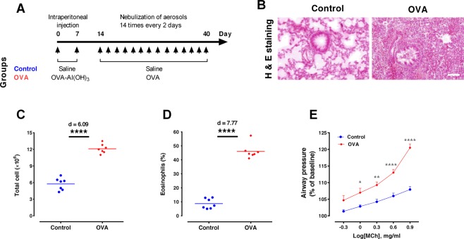
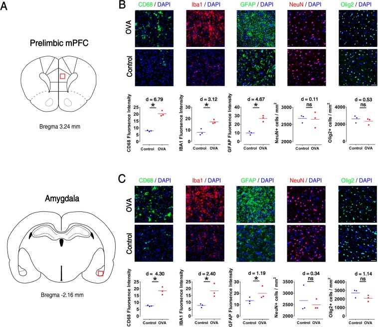
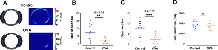
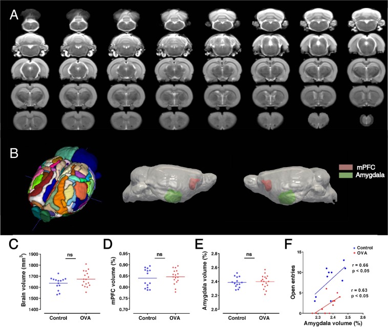
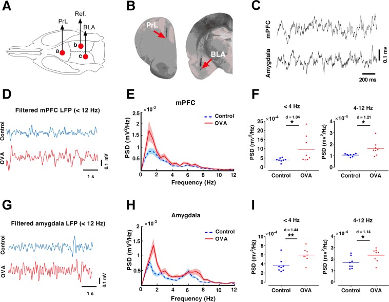
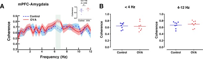
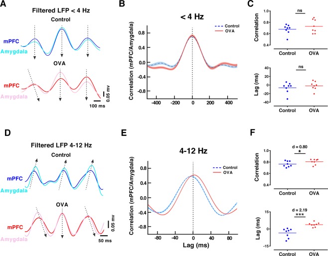
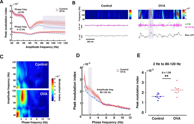
Anxiety is prevalent in asthma, and is associated with disease severity and poor quality of life. However, no study to date provides direct experimental evidence for the effect of allergic inflammation on the structure and function of medial prefrontal cortex (mPFC) and amygdala, which are essential regions for modulating anxiety and its behavioral expression. We assessed the impact of ovalbumin (OVA)-induced allergic inflammation on the appearance of anxiety-like behavior, mPFC and amygdala volumes using MRI, and the mPFC-amygdala circuit activity in sensitized rats. Our findings exhibited that the OVA challenge in sensitized rats induced anxiety-like behavior, and led to more activated microglia and astrocytes in the mPFC and amygdala. We also found a negative correlation between anxiety-like behavior and amygdala volume. Moreover, OVA challenge in sensitized rats was associated with increases in mPFC and amygdala activity, elevation of amygdala delta-gamma coupling, and the enhancement of functional connectivity within mPFC-amygdala circuit - accompanied by an inverted direction of information transferred from the amygdala to the mPFC. We indicated that disrupting the dynamic interactions of the mPFC-amygdala circuit may contribute to the induction of anxiety-related behaviors with asthma. These findings could provide new insight to clarify the underlying mechanisms of allergic inflammation-induced psychiatric disorders related to asthma.


Conflict of interest statement

The authors declare no competing interests.

Figures

Figure 1
Figure 1
Allergen exposure increases airway hyper-responsiveness and lung inflammation. (A) Timeline of the study design. The rats received 2 intraperitoneal injections of saline or OVA-Al(OH)3 [1 mg OVA (Grade III; Sigma) and 100 mg Al(OH)3, in 1 ml saline] on day 0 and day 7, and 14 inhalation exposures (for 30 min, every 2 days, from day 14 to day 40) with aerosols of saline or OVA solution (2% wt/vol). (B) Hematoxylin and eosin (H&E) staining of lung sections to identify peribronchial inflammatory cell infiltration (Scale bar = 50 μm). (C) Airway hyper-responsiveness in response to increasing doses of methacholine. Data are presented as percent of baseline, expressed as mean ± SEM, and analyzed by repeated measures two-way ANOVA, with the Bonferroni post-hoc test, n = 7 per group. (D,E) Total BAL inflammatory cells and eosinophils, respectively. The horizontal bars represent mean values. Data were analyzed by t-test, n = 7 per group. *p < 0.05, ***p < 0.001 and ****p < 0.0001 compared to control. OVA, ovalbumin.
Figure 2
Figure 2
Microglia and astrocytes were activated and oligodendrocytes were reduced in prelimbic mPFC and basolateral amygdala of allergen-exposured rats. (A) Displays schematics indicate regions of immunostaining in the brain. (B,C) Representative immunoreactivity images of microgelia (CD68 and Iba1), astrocyte (GFAP), neuronal survival (NeuN) and oligodendrocyte (Olig2) in prelimbic mPFC (B) and basolateral amygdala (C) as well as quantitative assessment on each test (lower panels). Scale bar represents 50 μm. Data were analyzed by Mann–Whitney test, n = 3 per group. *p < 0.05 compared to control. OVA, ovalbumin; mPFC, medical prefrontal cortex.
Figure 3
Figure 3
Allergen exposure induces anxiety-like behavior. (A) Representative animal track in the elevated zero maze for a control (upper panels) and an OVA (lower panels) rat. (B,C) Time spent in open sections and number of entries to the open sections in the elevated zero maze, which inversely related to anxiety-like behavior respectively (Control: n = 14, OVA: n = 11). (D) Total distance traveled (as an indicator of locomotor activity) in the open field test (Control: n = 13, OVA: n = 12). The horizontal bars represent mean values. Data were analyzed by t-test. **p < 0.01, ***p < 0.001 compared to control. ns, not significant; OVA, ovalbumin.
Figure 4
Figure 4
Anxiety-like behavior is negatively correlated with amygdala volume. (A) Representative T2-weighted coronal MRI slices, which were registered to template. (B) Displays three-dimensional rendering views of atlas (left panel), lateral representative of brain (middle and right panel). Regional selections of mPFC and amygdala have shown red and green, respectively. (CE) MRI volumetric measurements of whole brain, mPFC and amygdala, respectively. The horizontal bars represent mean values (as percent of brain volume). Data were analyzed by t-test (Control: n = 14, OVA: n = 17). (F) The number of entries to the open sections in elevated zero maze (inversely related to anxiety) was positively correlated with amygdala volume. ns, not significant; OVA, ovalbumin; mPFC, medical prefrontal cortex.
Figure 5
Figure 5
Allergen exposure increases delta and theta activity in mPFC and amygdala. (A) Schematic representation of electrode implantation sites. (B) Example histology of electrodes placed in PrL and BLA. (C) Representative raw traces of simultaneous LFP recordings from the mPFC and amygdala. (D,G) Examples of mPFC and amygdala LFP traces filtered at delta and theta frequencies (<12 Hz). (E,H) Averaged power spectra of LFPs in the mPFC and amygdala. Shaded area indicates standard errors. (F,I) OVA exposure increases delta (<4 Hz) and theta (4–12 Hz) power spectral density of the mPFC and amygdala LFPs. The horizontal bars represent mean values. Data were analyzed by t-test, n = 8 per group. *p < 0.05, **p < 0.01 compared to control. OVA, ovalbumin; mPFC, medical prefrontal cortex; PrL, prelimbic; BLA, basolateral amygdala; LFP, local field potential.
Figure 6
Figure 6
Coherence between mPFC and amygdala is increased at theta frequency in allergen-exposured rats. (A) Spectral coherence between simultaneously recorded mPFC and amygdala LFPs. Shaded area indicates standard errors. The gray area indicates significant differences between OVA and control rats. Inserted panel shows significant differences between OVA and control rats at 6.5–7 Hz frequencies. (B) Mean mPFC-amygdala coherence at delta (<4 Hz, left) and theta (4–12 Hz, right) frequencies. The horizontal bars represent mean values. Data were analyzed by t-test, n = 8 per group. *p < 0.05 compared to control. OVA, ovalbumin; mPFC, medical prefrontal cortex; LFP, local field potential.
Figure 7
Figure 7
Allergen exposure enhances synchrony and inverses directionality of theta activity in mPFC-amygdala circuit. (A,D) Examples of mPFC and amygdala delta (<4 Hz) and theta (4–12 Hz) filtered signals, illustrating the cross-correlation lag analysis in the control (upper) and OVA (lower). Arrows are drawn from the leading area to the lagging area. (B,E) Mean waveform correlation for the delta and theta-filtered signals in the mPFC-amygdala circuit. Shaded area indicates standard errors. (C,F) Mean mPFC-amygdala correlation and lag at delta (<4 Hz) and theta (4–12 Hz) frequencies. OVA exposure increases correlation for theta oscillation between mPFC and amygdala with a delay in the mPFC. The horizontal bars represent mean values. Data were analyzed by t-test, n = 8 per group. *p < 0.05, ***p < 0.001 compared to control. OVA, ovalbumin; mPFC, medical prefrontal cortex; LFP, local field potential.
Figure 8
Figure 8
Allergen exposure increases phase-amplitude coupling of delta and gamma oscillations in amygdala. (A) Peak modulation index across amplitude frequencies computed for delta (<4 Hz) and theta phase (4–12 Hz) in amygdala. Shaded area indicates standard errors. The gray area indicates significant differences between OVA and control rats. Delta modulates 80–120 gamma activity in OVA-exposured rats. (B) Spectrograms (color plots) of amygdala LFP (black, lower panels) and its delta (<4 Hz, green) and gamma (80–120 Hz, pink) filtered versions (middle panels) in control (left panels) and OVA (right panels) rats. Gray area indicates a representative high-amplitude gamma event occurring at the crest of delta phase. (C) Phase-amplitude comodugram of representative amygdala LFP recordings. (D) Peak modulation index across phase frequencies computed for gamma amplitude (80–120 Hz) in amygdala. Shaded area indicates standard errors. The gray area indicates significant differences between OVA and control rats. (E) Delta modulation of gamma increases for 2 Hz in OVA-exposured rats. The horizontal bars represent mean values. Data were analyzed by t-test, n = 8 per group. **p < 0.01 compared to control. OVA, ovalbumin; LFP, local field potential.

Similar articles

Cited by

References

    1. Strine TW, Mokdad AH, Balluz LS, Berry JT, Gonzalez O. Impact of depression and anxiety on quality of life, health behaviors, and asthma control among adults in the United States with asthma, 2006. J. Asthma. 2008;45:123–133. doi: 10.1080/02770900701840238. - DOI - PubMed
    1. Dudeney J, Sharpe L, Jaffe A, Jones EB, Hunt C. Anxiety in youth with asthma: A meta-analysis. Pediatr. Pulmonol. 2017;52:1121–1129. doi: 10.1002/ppul.23689. - DOI - PubMed
    1. Rosenkranz MA, et al. From The Cover: Neural circuitry underlying the interaction between emotion and asthma symptom exacerbation. Proc. Natl. Acad. Sci. 2005;102:13319–13324. doi: 10.1073/pnas.0504365102. - DOI - PMC - PubMed
    1. Costa-Pinto FA, Basso AS, Britto LRG, Malucelli BE, Russo M. Avoidance behavior and neural correlates of allergen exposure in a murine model of asthma. Brain. Behav. Immun. 2005;19:52–60. doi: 10.1016/j.bbi.2004.02.005. - DOI - PubMed
    1. Palermo-Neto J, Guimarães RK. Pavlovian conditioning of lung anaphylactic response in rats. Life Sci. 2000;68:611–623. doi: 10.1016/S0024-3205(00)00966-8. - DOI - PubMed

Publication types