. 2019 Dec 20;9(1):19565.
doi: 10.1038/s41598-019-56021-w.

Controlling properties of human neural progenitor cells using 2D and 3D conductive polymer scaffolds

Affiliations

Controlling properties of human neural progenitor cells using 2D and 3D conductive polymer scaffolds

Shang Song et al. Sci Rep. .

Abstract

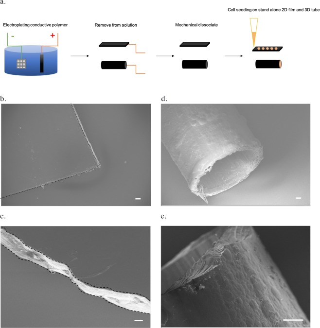
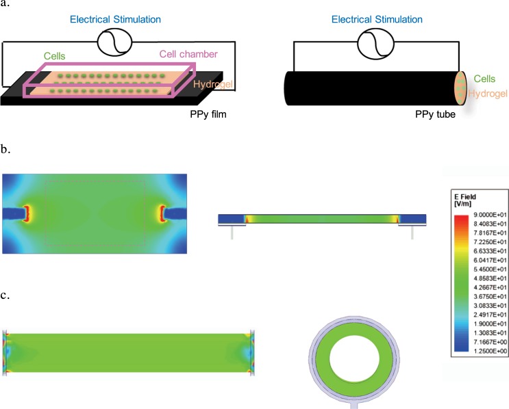
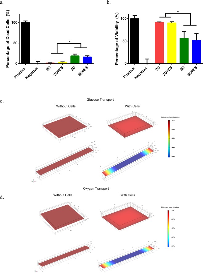
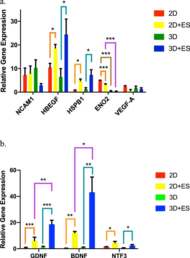
Human induced pluripotent stem cell-derived neural progenitor cells (hNPCs) are a promising cell source for stem cell transplantation to treat neurological diseases such as stroke and peripheral nerve injuries. However, there have been limited studies investigating how the dimensionality of the physical and electrical microenvironment affects hNPC function. In this study, we report the fabrication of two- and three-dimensional (2D and 3D respectively) constructs composed of a conductive polymer to compare the effect of electrical stimulation of hydrogel-immobilized hNPCs. The physical dimension (2D vs 3D) of stimulating platforms alone changed the hNPCs gene expression related to cell proliferation and metabolic pathways. The addition of electrical stimulation was critical in upregulating gene expression of neurotrophic factors that are important in regulating cell survival, synaptic remodeling, and nerve regeneration. This study demonstrates that the applied electrical field controls hNPC properties depending on the physical nature of stimulating platforms and cellular metabolic states. The ability to control hNPC functions can be beneficial in understanding mechanistic changes related to electrical modulation and devising novel treatment methods for neurological diseases.


Conflict of interest statement

There are no competing or conflicts of interest related to the work presented in this manuscript for the authors.

Figures

Figure 1
Figure 1
Fabrication and structures of the conductive polypyrrole (PPy) scaffolds. (a) Electroplating of the PPy scaffold (black) using platinum mesh (grey) as the reference under applied voltage (green and red as source electrodes) in the NaDBS-doped PPy solution (blue). The PPy scaffolds were mechanically dissociated into 2D films sand 3D tubes, where cells (orange) were deposited on top or inside, respectively. (b) Scanning Electron Microscopy (SEM) image showing the top view of the 2D conductive PPy film (outlined in dashes, scale bar: 100 μm). (c) The cross-section SEM image of the 2D PPy film (outlined in dashes, scale bar: 20 μm). (d) The hollow 3D conductive PPy tube (scale bar: 100μm). (e) The side view of sectioned 3D PPy tube (scale bar: 100 μm).
Figure 2
Figure 2
The electrically-stimulated hNPC-seeded polypyrrole (PPy) scaffolds. (a) The longitudinal ends of the 2D PPy film (left) or 3D PPy tube (right) were attached with external wires for electrical stimulation. The hydrogel-immobilized cells (green) were seeded directly on top of the 2D PPy film within the cell chamber or encapsulated inside the 3D PPy tube. The Electromagnetic field computation with ANSYS HFSS showed the distribution of the electrical field under applied electrical stimulation in (b) the top and side views of 2D PPy film with dashline showing where cells were seeded, and (c) the top and cross-section views of 3D PPy tube. The computational modeling indicated that the electrical field was the strongest at the point of contact with the external electrical sources and the electrical fields were uniform across scaffolds with similar field strength (~40 V/m) between the 2D and 3D PPy scaffolds.
Figure 3
Figure 3
The viability of electrically stimulated hNPCs in the 2D and 3D conductive PPy scaffolds. (a) The lactate dehydrogenase (LDH) assay showed no significant difference in the percentage of dead cells caused by electrical stimulation in both unstimulated (2D) and stimulated (2D + ES) 2D PPy films, and unstimulated (3D) and stimulated (3D + ES) 3D PPy tubes, respectively. However, cells encapsulated in the 3D PPy tubes showed significantly higher cell death compared to all 2D conditions. (b) The alamar blue assay showed that the viability was similar between the unstimulated (2D) and stimulated 2D PPy films, and the unstimulated (3D) and stimulated (3D) PPy tubes, respectively. In general, all 2D conditions showed better cell viability compared to the 3D conditions. (n = 4, error bars show SE, *p < 0.05) Nutrient molecule transport with COMSOL Multiphysics showed (c) glucose and (d) oxygen gradient in the 2D PPy film (top) and 3D PPy tube (bottom). With cells (right panels), there were significant changes in nutrient exchange for both scaffolds. The 3D PPy tube showed a significant change in glucose and oxygen concentrations within the tube compared to the 2D PPy film.
Figure 4
Figure 4
Gene expression changes with electrical stimulation using 2D and 3D conductive PPy scaffolds. The fold change in the relative gene expression of (a) NCAM1, HBEGF, HSPB1, ENO2, and VEGF-A and (b) GDNF, BDNF, and NTF3 in unstimulated (2D) and stimulated hNPCs (2D + ES) on 2D PPy films and in unstimulated (3D) and stimulated hNPCs (3D + ES) in 3D PPy tubes. Orange lines indicated comparison between the “2D” and “2D + ES” groups. Teal lines indicated comparison between the “3D” and “3D + ES” groups. Brown lines showed comparison between the “2D” and “3D” groups. Purple lines showed comparison between the “2D + ES” and “3D + ES” groups. (n = 4, error bars show SE, One-way ANOVA followed by multiple comparisons by Tukey, *p ≤ 0.05, **p ≤ 0.01, ***p ≤ 0.001).

Similar articles

Cited by

References

    1. Kabu S, Gao Y, Kwon BK, Labhasetwar V. Drug delivery, cell-based therapies, and tissue engineering approaches for spinal cord injury. J Control Release. 2015;219:141–154. doi: 10.1016/j.jconrel.2015.08.060. - DOI - PMC - PubMed
    1. Menzies FM, Fleming A, Rubinsztein DC. Compromised autophagy and neurodegenerative diseases. Nature Reviews Neuroscience. 2015;16:345–357. doi: 10.1038/nrn3961. - DOI - PubMed
    1. Gorman AM. Neuronal cell death in neurodegenerative diseases: recurring themes around protein handling. J. Cell. Mol. Med. 2008;12:2263–2280. doi: 10.1111/j.1582-4934.2008.00402.x. - DOI - PMC - PubMed
    1. Haston KM, Finkbeiner S. Clinical Trials in a Dish: The Potential of Pluripotent Stem Cells to Develop Therapies for Neurodegenerative Diseases. Annu. Rev. Pharmacol. Toxicol. 2016;56:489–510. doi: 10.1146/annurev-pharmtox-010715-103548. - DOI - PMC - PubMed
    1. Trounson A, McDonald C. Stem Cell Therapies in Clinical Trials: Progress and Challenges. Cell Stem Cell. 2015;17:11–22. doi: 10.1016/j.stem.2015.06.007. - DOI - PubMed

Publication types