. 2020 Feb 28;48(4):1969-1984.
doi: 10.1093/nar/gkz1185.

Adapted formaldehyde gradient cross-linking protocol implicates human eIF3d and eIF3c, k and l subunits in the 43S and 48S pre-initiation complex assembly, respectively

Affiliations

Adapted formaldehyde gradient cross-linking protocol implicates human eIF3d and eIF3c, k and l subunits in the 43S and 48S pre-initiation complex assembly, respectively

Anna Herrmannová et al. Nucleic Acids Res. .

Abstract

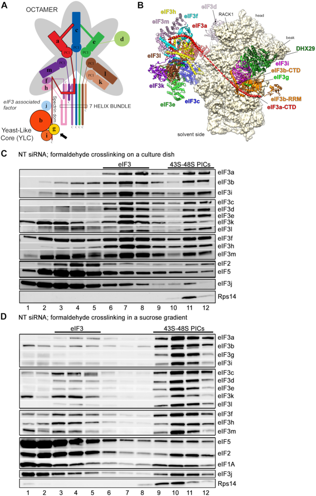
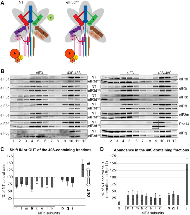
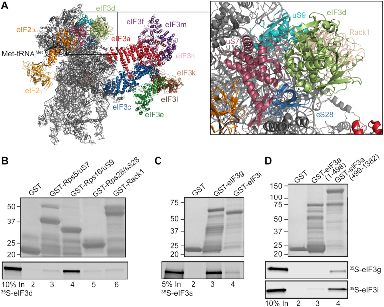
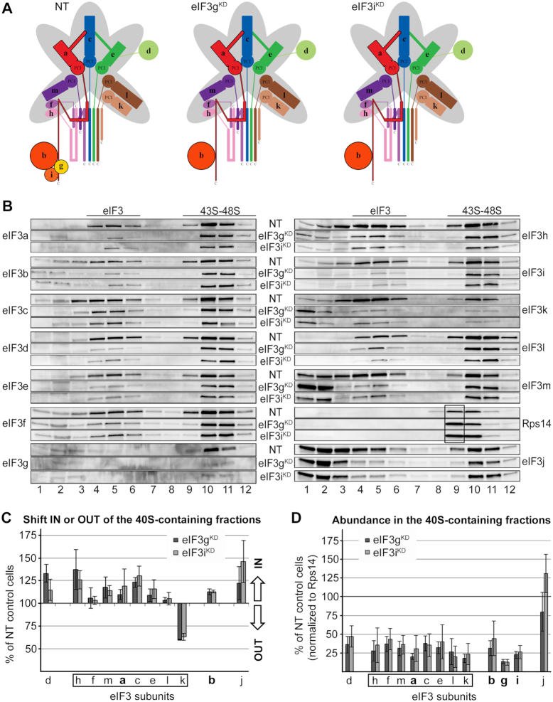
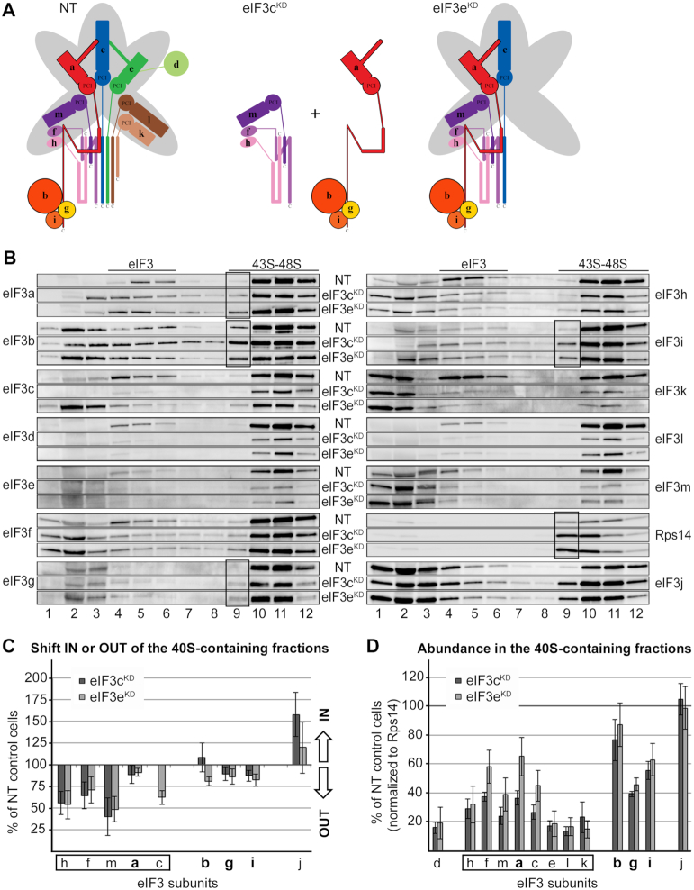
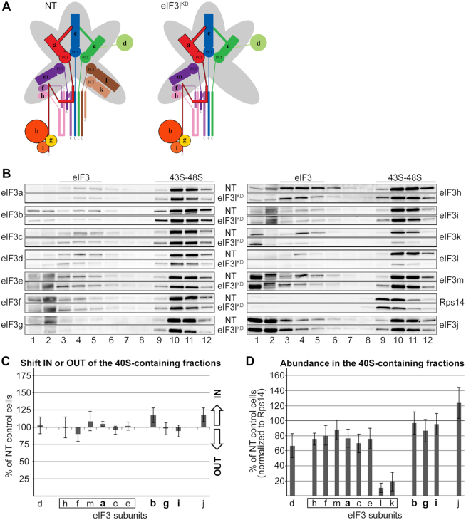
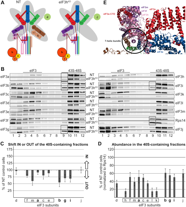
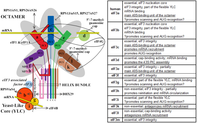
One of the key roles of the 12-subunit eukaryotic translation initiation factor 3 (eIF3) is to promote the formation of the 43S and 48S pre-initiation complexes (PICs). However, particular contributions of its individual subunits to these two critical initiation reactions remained obscure. Here, we adapted formaldehyde gradient cross-linking protocol to translation studies and investigated the efficiency of the 43S and 48S PIC assembly in knockdowns of individual subunits of human eIF3 known to produce various partial subcomplexes. We revealed that eIF3d constitutes an important intermolecular bridge between eIF3 and the 40S subunit as its elimination from the eIF3 holocomplex severely compromised the 43S PIC assembly. Similarly, subunits eIF3a, c and e were found to represent an important binding force driving eIF3 binding to the 40S subunit. In addition, we demonstrated that eIF3c, and eIF3k and l subunits alter the efficiency of mRNA recruitment to 43S PICs in an opposite manner. Whereas the eIF3c knockdown reduces it, downregulation of eIF3k or eIF3l increases mRNA recruitment, suggesting that the latter subunits possess a regulatory potential. Altogether this study provides new insights into the role of human eIF3 in the initial assembly steps of the translational machinery.


Figures

Figure 1.
Figure 1.
Comparison of the original ‘on dish’ formaldehyde cross-linking with the optimized formaldehyde cross-linking sucrose gradient protocol based on GraFix. (A) Schematic model of the human eIF3 complex adapted from (7). eIF3 subunits forming the PCI/MPN octamer are indicated by the grey background. The rectangle marks the seven α-helices involved in formation of the 7-helix bundle (5). The Yeast-Like Core (YLC) comprising the eIF3 subunits a, b, g and i is depicted, and so is the eIF3-associated factor eIF3j. Arrow indicates newly identified contact between eIF3g and the eIF3a-CTD (see Figure 3C and D). (B) Model of the structure of the mammalian 43S pre-initiation complex adapted from (56). The 43S PIC is viewed from the solvent side. The red dashed line represents continuity of the eIF3a structure (its C-terminal tail) that remains unsolved. (C, D) The 43S PIC assembly analysis of the control NT cells prepared by (C) the ‘on dish’ formaldehyde crosslinking protocol (adapted from (8)) or (D) the optimized protocol developed in this study. Antibodies used for western blotting are indicated on the right side of each panel. Sucrose gradient fraction numbers are given at the bottom. Fraction 1 in (D) is a pool of the first two fractions from the top of the gradient. Fractions containing ribosome free versus bound eIF3 complexes (eIF3 versus 43S-48S PICs) are indicated at the top.
Figure 2.
Figure 2.
eIF3d critically promotes the 43S PIC formation. (A) Comparison of schematic models of eIF3 complexes occurring in the control NT versus eIF3dKD cells. (B) The 43S PIC assembly analysis of the control NT versus eIF3dKD cells performed as described in Materials and Methods. Please note that for easier comparison we coupled Western blot membrane strips corresponding to individual eIF3 subunits together with that coming from control NT cells always above the one derived from the eIF3dKD (follow the labeling in between the two panels). Sucrose gradient fraction numbers are given at the bottom. Fraction 1 in is a pool of the first two fractions from the top of the gradient. Fractions containing ribosome free versus bound eIF3 complexes (eIF3 versus 43S–48S PICs) are indicated at the top. These experiments were performed three times. For description of the black box, see the main text (C) Relative change in distribution of individual eIF3 subunits in the 40S containing fractions with respect to the entire gradient. Values >100% signal accumulation of a factor of interest in the 40S-containing fractions, whereas values <100% point to its redistribution to lighter fractions. For details, see Materials and Methods. Please note that the distribution across the gradient was calculated for non-downregulated subunits only. Octameric eIF3 subunits are highlighted by a rectangle, the YLC subunits in bold. (D) Relative amounts of individual eIF3 subunits bound per ribosome (normalized to the Rps14 amount) compared to the control NT cells. For details, see Materials and Methods. For both quantifications, the Quantity One software from Bio-Rad was used.
Figure 3.
Figure 3.
eIF3d interacts with Rps16/uS9 and the eIF3a-CTD binds both eIF3i and g subunits of the YLC in vitro. (A) A cryo-EM model of the eIF3d placement on the 40S head highlighting its prospective contacts with small ribosomal proteins (adapted from (46)). (B) eIF3d interacts with Rps16/uS9 in vitro. Small ribosomal proteins Rps5/uS7 (lane 3), Rps16/uS9 (lane 4), Rps28/eS28 (lane 5) and Rack1 (lane 6) fused to GST moiety, and GST alone (lane 2) were tested for binding to 35S-labeled eIF3d. Lane 1 (In) contains 10% of input amounts of radiolabeled eIF3d added to each reaction mixture. (C, D) The eIF3a-CTD binds both eIF3i and g subunits of the YLC in vitro. (C) The eIF3g (lane 3) and eIF3i (lane 4) subunits fused to GST moiety, and GST alone (lane 2) were tested for binding to 35S-labeled eIF3a. Lane 1 (In) contains 5% of input amounts of radiolabeled eIF3a added to each reaction mixture. (D) The eIF3a N-terminal fragment (1–498 aa) (lane 3) and the eIF3a C-terminal fragment (499–1382 aa) (lane 4) fused to GST moiety, and GST alone (lane 2) were tested for binding to 35S-labeled eIF3g and eIF3i. Lane 1 (In) contains 10% of input amounts of radiolabeled eIF3g or eIF3i added to each reaction mixture.
Figure 4.
Figure 4.
eIF3g and eIF3i are dispensable for the assembly of initiation complexes. (A) Comparison of schematic models of eIF3 complexes occurring in the control NT versus eIF3gKD and eIF3iKD cells. (BD) Same as in Figure 2B–D except that the eIF3gKD and eIF3iKD were analyzed. These experiments were performed four times.
Figure 5.
Figure 5.
The effect of eIF3c and eIF3e knockdowns on the 43S PIC assembly. (A) Comparison of schematic models of eIF3 complexes occurring in the control NT versus eIF3cKD and eIF3eKD cells. (BD) Same as in Figure 2B–D except that the eIF3cKD and eIF3eKD were analyzed. These experiments were performed six times. Black boxes indicate the occurrence of the YLC (a-b-g-i) in fraction 9.
Figure 6.
Figure 6.
The eIF3lKD does not affect association of eIF3 with 40S ribosomes. (A) Comparison of schematic models of eIF3 complexes occurring in the control NT versus eIF3lKD cells. (BD) Same as in Figure 2B–D except that the eIF3lKD was analyzed. These experiments were performed six times.
Figure 7.
Figure 7.
The eIF3hKD impairs accommodation of the eIF3 b-g-i module on the 40S subunit. (A) Comparison of schematic models of eIF3 complexes occurring in the control NT versus eIF3hKD cells. The eIF3 subunits with decreased protein levels are underlined in the eIF3hKD schematic (BD) Same as in Figure 2B–D except that the eIF3hKD was analyzed. These experiments were performed six times. Black boxes indicate the occurrence of the YLC (a-b-g-i) in fraction 9. (E) A cryo-EM model of the 7-helix bundle with the eIF3f-h-m module on top of it being wrapped around by the initial segment of the eIF3a C-terminal tail (adapted from (46)).
Figure 8.
Figure 8.
A schematic model of human eIF3 (adapted from (2)), with a table summarizing functional contributions of all individual human eIF3 subunits to general translation initiation known to date from our work and the work of our colleagues (1–3,7–11,21,22,28–31); findings from this work are underlined. The eIF3 subunits forming the PCI/MPN octamer are indicated by the grey background. The rectangle marks the seven α-helices involved in formation of the 7-helix bundle; the Yeast-Like Core (YLC) comprising the eIF3 subunits a, b, g and i is depicted at the bottom. Arrows indicate all known interactions of eIF3 subunits with other eIFs, ribosomal proteins and mRNA (reviewed in (2)).

Similar articles

Cited by

References

    1. Gomes-Duarte A., Lacerda R., Menezes J., Romão L.. eIF3: a factor for human health and disease. RNA Biol. 2018; 15:26–34. - PMC - PubMed
    1. Valášek L.S., Zeman J., Wagner S., Beznosková P., Pavlíková Z., Mohammad M.P., Hronová V., Herrmannová A., Hashem Y., Gunišová S.. Embraced by eIF3: structural and functional insights into the roles of eIF3 across the translation cycle. Nucleic Acids Res. 2017; 45:10948–10968. - PMC - PubMed
    1. Cate J.H.D. Human eIF3: from ‘blobology’ to biological insight. Philos. Trans. R. Soc. Lond. B. Biol. Sci. 2017; 372:20160176. - PMC - PubMed
    1. Querol-Audi J., Sun C., Vogan J.M., Smith M.D., Gu Y., Cate J.H.D., Nogales E.. Architecture of human translation initiation factor 3. Structure. 2013; 21:920–928. - PMC - PubMed
    1. des Georges A., Dhote V., Kuhn L., Hellen C.U.T., Pestova T.V, Frank J., Hashem Y.. Structure of mammalian eIF3 in the context of the 43S preinitiation complex. Nature. 2015; 525:491–495. - PMC - PubMed

Publication types

MeSH terms