Review
. 2020 Mar 6;432(5):1310-1325.
doi: 10.1016/j.jmb.2019.12.012. Epub 2019 Dec 19.

Recent Insights into Beta-cell Exocytosis in Type 2 Diabetes

Affiliations
Review

Recent Insights into Beta-cell Exocytosis in Type 2 Diabetes

Debbie C Thurmond et al. J Mol Biol. .

Abstract

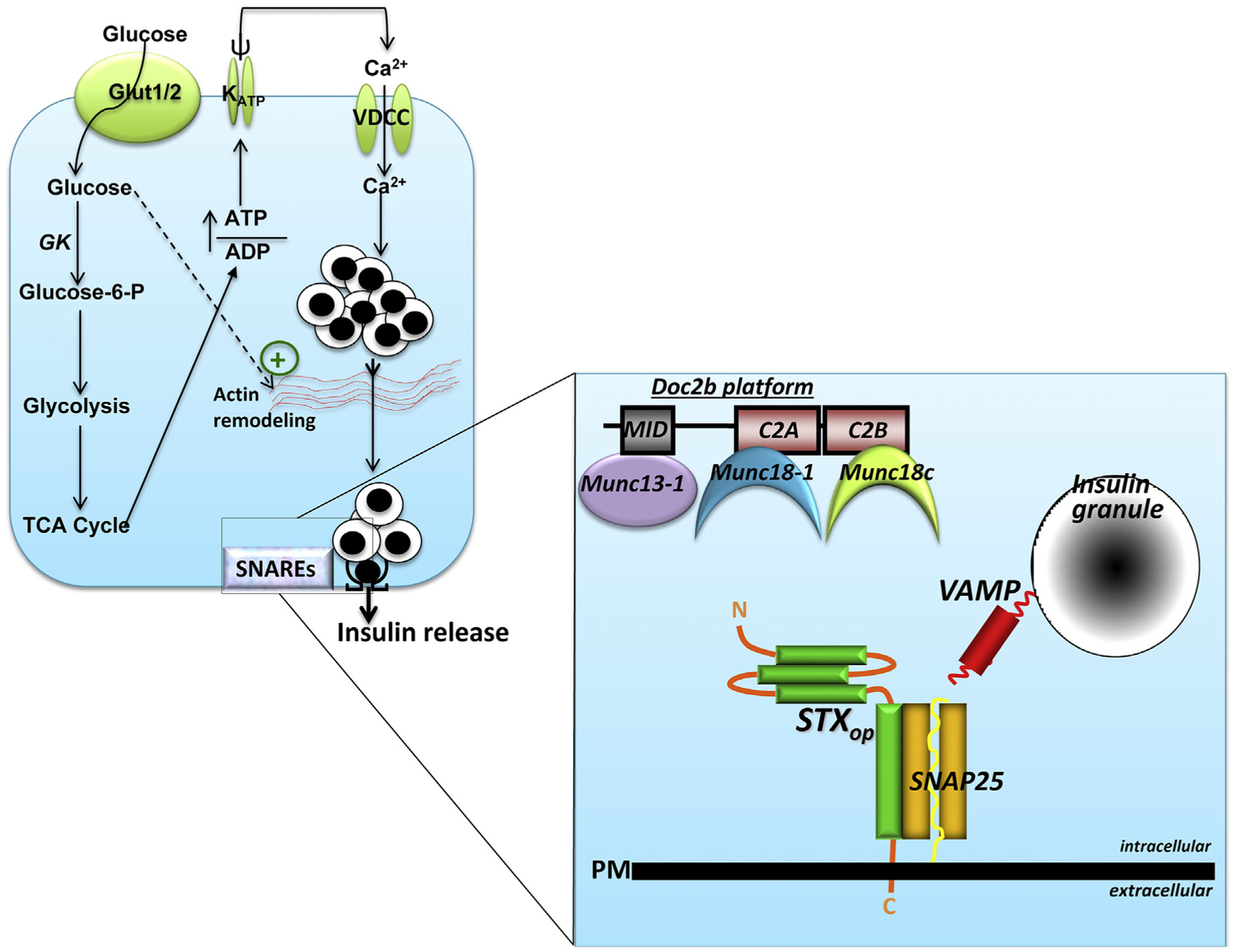
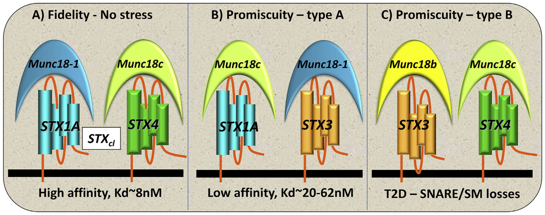
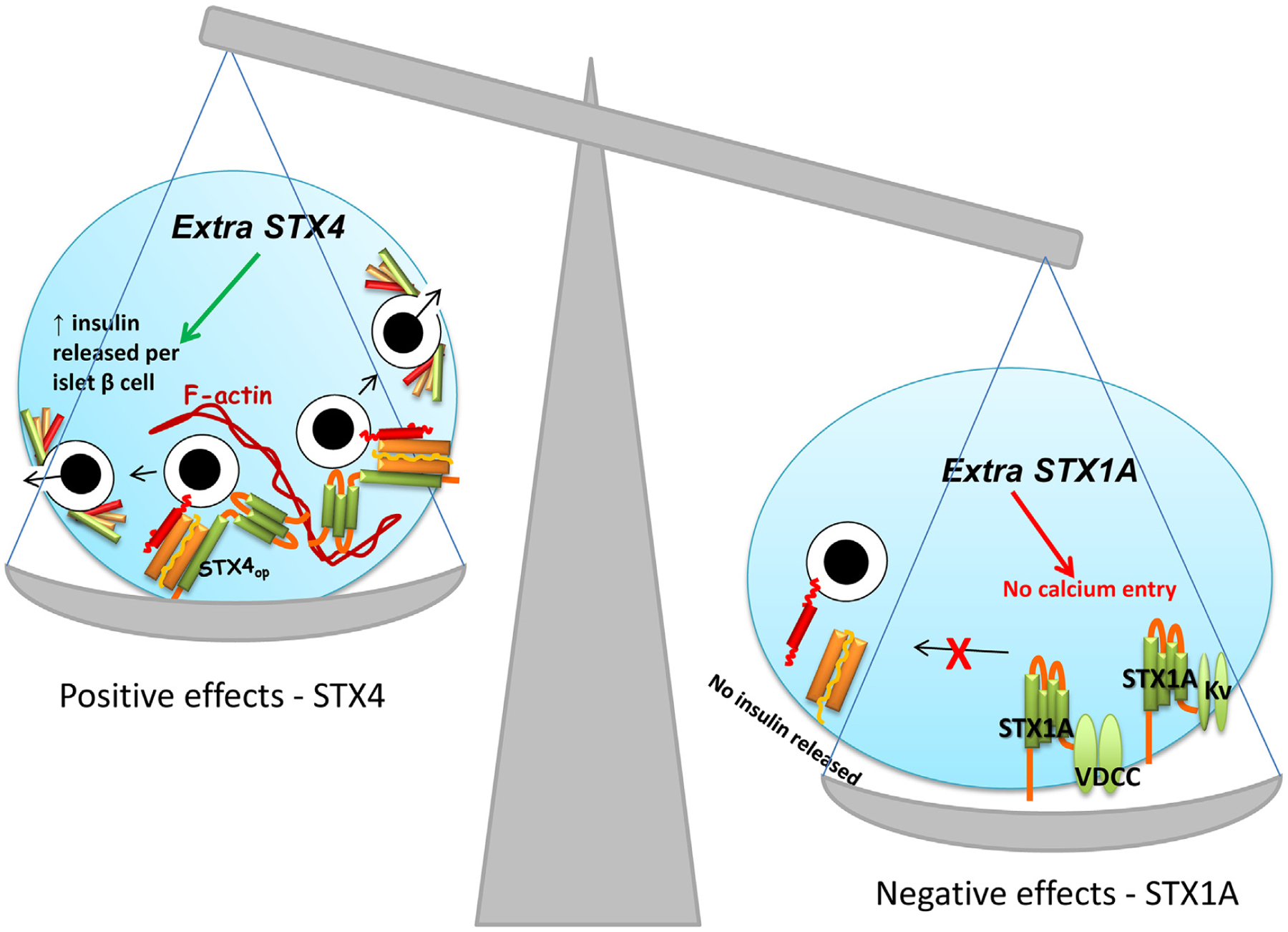
As one of the leading causes of morbidity and mortality worldwide, diabetes affects an estimated 422 million adults, and it is expected to continue expanding such that by 2050, 30% of the U.S. population will become diabetic within their lifetime. Out of the estimated 422 million people currently afflicted with diabetes worldwide, about 5% have type 1 diabetes (T1D), while the remaining ~95% of diabetics have type 2 diabetes (T2D). Type 1 diabetes results from the autoimmune-mediated destruction of functional β-cell mass, whereas T2D results from combinatorial defects in functional β-cell mass plus peripheral glucose uptake. Both types of diabetes are now believed to be preceded by β-cell dysfunction. T2D is increasingly associated with numerous reports of deficiencies in the exocytosis proteins that regulate insulin release from β-cells, specifically the soluble N-ethylmaleimide-sensitive factor attachment protein receptor (SNARE) proteins. SNARE protein's functionality is further regulated by a variety of accessory factors such as Sec1/Munc18 (SM), double C2-domain proteins (DOC2), and additional interacting proteins at the cell surface that influence the fidelity of insulin release. As new evidence emerges about the detailed mechanisms of exocytosis, new questions and controversies have come to light. This emerging information is also contributing to dialogue in the islet biology field focused on how to correct the defects in insulin exocytosis. Herein we present a balanced review of the role of exocytosis proteins in T2D, with thoughts on novel strategies to protect functional β-cell mass.

Keywords: SM proteins; SNARE proteins; glucose-stimulated insulin secretion; insulin granule; islet beta cell.


Figures

Fig. 1.
Fig. 1.. Type 2 diabetic (T2D) human islets show deficient first- and second-phase glucose-stimulated insulin release (GSIS).
Biphasic insulin release in response to a glucose stimulus measured in isolated human islets from age, gender, and ethnicity matched human donors who are nondiabetic (red line) or T2D (black line). First-phase release begins within 2 min and lasts for ~10 min (yellow region), whereas second-phase release can last as long as several hours, if the glucose stimulus persists (orange region). Note that first phase does not return to baseline when the second phase begins. Adapted from Ref. [10], with permission from JCEM.
Fig. 2.
Fig. 2.. Basic steps in insulin granule exocytosis.
β-cells sense elevated circulating glucose via the glucose transporter (Glut1 in humans, Glut2 in rodents) at the plasma membrane (PM). Glucose is rapidly metabolized, which increases the ATP/ADP ratio, triggering KATP channel closure, PM depolarization (ψ), and opening of the voltage-dependent calcium channels (VDCC) to increase intracellular calcium. Glucose also promotes actin remodeling to facilitate the steady flow of insulin granules to the SNAREs at the PM. The details of the SNAREs are shown in an enlargement at the right, where the three SNAREs (STXop, open form of syntaxin; SNAP25, and VAMP) are assembling to form the heterotrimeric SNARE complex. After SNARE complex formation, the ISGs fuse with the PM and the insulin cargo is released into the extracellular space. The SNARE complex assembly process is regulated by accessory proteins that can attach to a scaffold protein, DOC2B. The DOC2B scaffold acts as a floating platform so that when the Munc18 proteins release from their cognate STX partners, they can associate with the nearby Doc2b platform. Munc18–1 binds to Doc2b′s C2A domain, and Munc18c binds to Doc2b′s C2B domain; the priming protein Munc13–1 can associate with Doc2b via its Munc13-interacting domain (MID).
Fig. 3.
Fig. 3.. Fidelity and promiscuity in β-cell SNARE pairing.
(A) Fidelity occurs in healthy β-cells, with STX and Munc18 isoforms pairing according to highest affinity partnerships (Kd ~8 nM). (B) Promiscuity type A occurs under conditions of stress where lower-affinity partnerships are formed (Kd ~20–62 nM) and GSIS function is reduced. (C) Promiscuity type B occurs in the absence or greatly reduced amount of a primary SNARE (e.g., STX1A) or Munc18 isoform (e.g., Munc18–1) in T2D islets, which can be overcome by the overexpression of Munc18b or STX4, resulting in functionally redundant rescue of those deficient exocytotic proteins.
Fig. 4.
Fig. 4.. Functional and dysfunctional responses to disruptions in stoichiometry.
(A) An overabundance of STX4 in the β-cell increases the amplitude of both phases of GSIS and enhances glucose homeostasis in vivo. STX4 binds directly to F-actin and indirectly via association with the actin binding protein gelsolin. Additional units of STX4 increase the abundance of STX4-based SNARE complexes. (B) An overabundance of STX1A decreases VDCC and Kv channel activity/gating, linked to STX1A’s ability to bind to the channels and reduce function, which in vivo decreases GSIS and disrupts glucose homeostasis.

References

    1. Curry DL, Bennett LL, Grodsky GM, Dynamics of insulin secretion by the perfused rat pancreas, Endocrinology 83 (1968) 572–584. - PubMed
    1. Porte D Jr., Pupo AA Insulin responses to glucose: evidence for a two pool system in man, J. Clin. Investig 48 (1969) 2309–2319. - PMC - PubMed
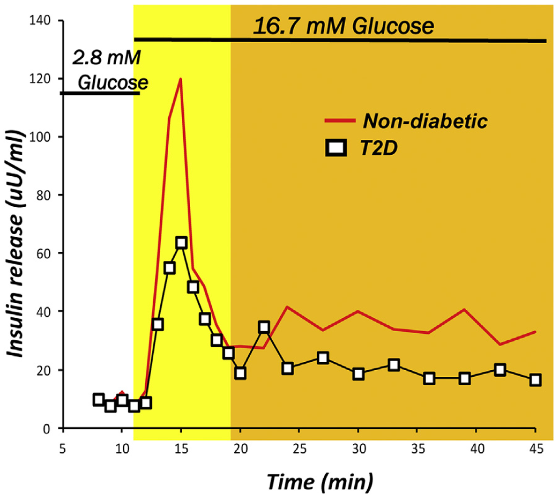
    1. Daniel S, Noda M, Straub SG, Sharp GW, Komatsu M, Schermerhorn T, et al., Identification of the docked granule pool responsible for the first phase of glucose-stimulated insulin secretion, Diabetes 48 (1999) 1686–1690. - PubMed
    1. Barg S, Eliasson L, Renstrom E, Rorsman P, A subset of 50 secretory granules in close contact with L-type Ca(2+) channels accounts for first-phase insulin secretion in mouse beta-cells, Diabetes 51 (2002) S74–S82. - PubMed
    1. Olofsson CS, Gopel SO, Barg S, Galvanovskis J, Ma X, Salehi A, et al., Fast insulin secretion reflects exocytosis of docked granules in mouse pancreatic B-cells, Pflüg. Arch 444 (2002) 43–51. - PubMed

Publication types