. 2019 Dec 21;19(1):333.
doi: 10.1186/s12883-019-1572-3.

The eagle jugular syndrome

Affiliations

The eagle jugular syndrome

Paolo Zamboni et al. BMC Neurol. .

Abstract

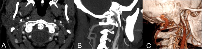
Background: The elongation of the styloid process is historically associated with two variants of the Eagle syndrome. The classic one, mainly characterized by pain and dysphagia, and the carotid variant characterized by pain and sometimes by cerebral ischemia. We observed a further variant characterized by a styloid elongation coursing adjacent to the transverse process of C1, causing significant compression of the internal jugular vein.

Methods: We reviewed all the cases of Eagle syndrome, including the jugular variant, admitted in our Hospital in the last six years. We compared symptomatology, associated comorbidities and imaging. Data were statistically analyzed.

Results: Overall 23 patients were admitted to the Hospital for symptomatic elongation of the styloid process, 11 male and 12 females. The jugular variant of the Eagle syndrome is clinically delineated by significant differences, as compared to the classic variant and carotid variants. Headache was the more prominent symptom (p < .009) as well as a documented peri-mesencephalic hemorrhage was the more significant comorbidity (p < .0003). The group classic-carotid variant was characterized by ipsilateral pain respect to the jugular variant (p < .0003). CT angiography with venous phase extended to the neck veins and imaging reconstruction is highly recommended as imaging technique, complemented by color-Doppler ultrasound.

Conclusions: The elongation of the styloid process may have different paths which creates compression on the surrounding anatomical structures. There may be a possible association of jugular impingement by an elongated styloid process with symptoms.

Trial registration: Protocol n°45-2013.

Keywords: Eagle syndrome; Elongated styloid process; Jugular compression; Perimesencephalic subarachnoid haemorrhage.


Conflict of interest statement

The authors declare that they have no competing interests.

Figures

Fig. 1
Fig. 1
CT angio of the illustrative case showing a right jugular internal vein bone nutcracker between the C1 transverse process and the elongated styloid process. a) Axial cut; b) Longitudinal reconstruction; c) 3D reconstruction
Fig. 2
Fig. 2
Transverse access CDU of the upper neck of the illustrative case. It is well apparent on the left a collapsed empty internal jugular vein (IJV) without any Doppler flow signal and an enlarged external jugular vein (EJV). CCA: common carotid artery. Right: six months post operatory CDU of the illustrative case, at the level of the carotid bifurcation. A filled and expanded IJV with Doppler flow signal is well apparent. ICA: internal carotid artery. ECA: external carotid artery
Fig. 3
Fig. 3
a) intraoperative picture of illustrative case, showing the relationship between the elongated styloid process (°) and the internal jugular vein (*); b) the elongation of the removed styloid process; c) the post-operative CTA showing the obtained decompression of the jugular vein due to the styloid process removal, in the absence of the restoration of its flow. This lead us to perform the following jugular PTA
Fig. 4
Fig. 4
a) Catheter venography of the right internal jugular vein showing the significant reduction of the cross sectional area either at the upper level, corresponding to the previous site of compression, or at the lower level, corresponding to a rigid valve apparatus; b-c). Delayed clearance of the contrast dye; d) Optimal morphological result after balloon angioplasty (PTA), functionally corresponding to a prompt drainage of the contrast dye
Fig. 5
Fig. 5
CTA showing a classic Eagle syndrome. The arrow points out the elongated styloid process. The space between this and the transverse process of C1, where the Jugular Vein courses, is wider than the jugular variant

References

    1. Fusco DJ, Asteraki S, Spetzler RF. Eagle's syndrome: embryology, anatomy, and clinical management. Acta Neurochir. 2012;154(7):1119–1126. doi: 10.1007/s00701-012-1385-2. - DOI - PubMed
    1. Babad MS. Eagle's syndrome caused by traumatic fracture of a mineralized stylohyoid ligament--literature review and a case report. Cranio. 1995;13(3):188–192. doi: 10.1080/08869634.1995.11678067. - DOI - PubMed
    1. Pierrakou ED. Eagle's syndrome. Review of the literature and a case report. Ann Dent. 1990;49(1):30–33. - PubMed
    1. Han MK, Kim DW, Yang JY. Non surgical treatment of Eagle's syndrome - a case report. Korean J Pain. 2013;26(2):169–172. doi: 10.3344/kjp.2013.26.2.169. - DOI - PMC - PubMed
    1. Politi M, Toro C, Tenani G. A rare cause for cervical pain: Eagle's syndrome. Int J Dent. 2009;2009:781297. doi: 10.1155/2009/781297. - DOI - PMC - PubMed

Supplementary concepts

LinkOut - more resources