. 2019 Dec 21;19(1):264.
doi: 10.1186/s12886-019-1265-0.

Risk factors for glial cell proliferation after idiopathic macular hole repair with internal limiting membrane flap

Affiliations

Risk factors for glial cell proliferation after idiopathic macular hole repair with internal limiting membrane flap

Yuyan Liu et al. BMC Ophthalmol. .

Abstract

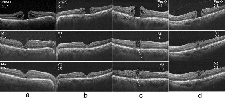
Background: To study the influencing factors for different healing patterns of patients with idiopathic macular holes (IMH) after vitrectomy surgery performed with the internal limiting membrane (ILM) flap technique.

Methods: This study was a retrospective, consecutive, observational case series study. We recruited 52 IMH patients who underwent vitrectomy with the ILM flap technique. The participants were divided into 2 groups: group A (25 patients), without significant glial cell proliferation in the macular area on postoperative optical coherence tomography (OCT); and group B (27 patients), with significant glial cell proliferation. The postoperative visual acuity (VA), external limiting membrane (ELM) and ellipsoid zone (EZ) recovery characteristics were compared between the two groups.

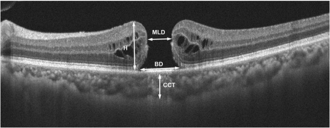
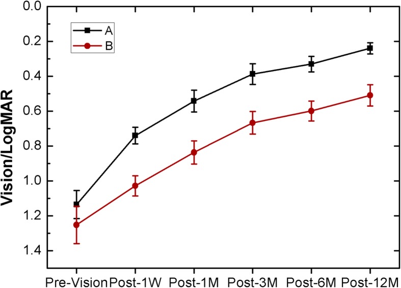
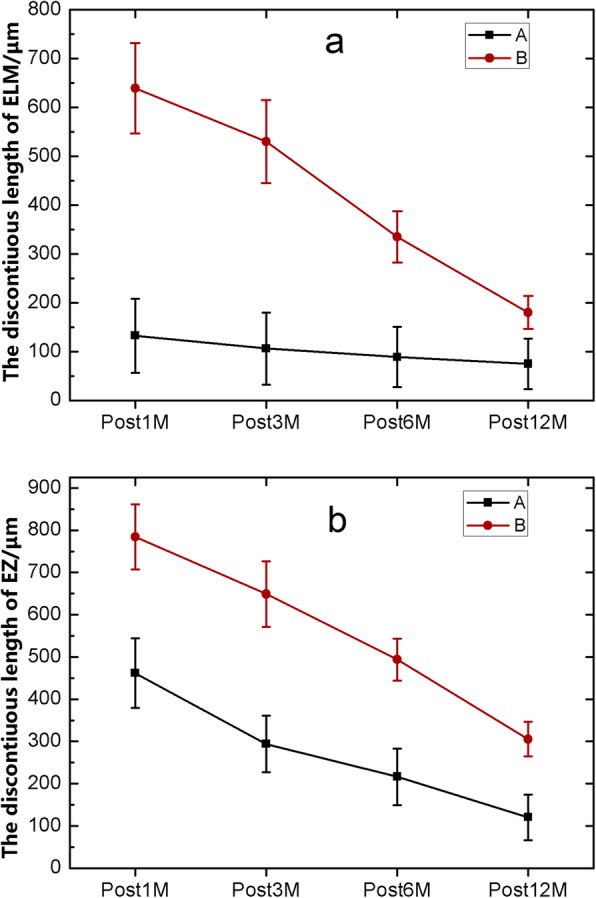
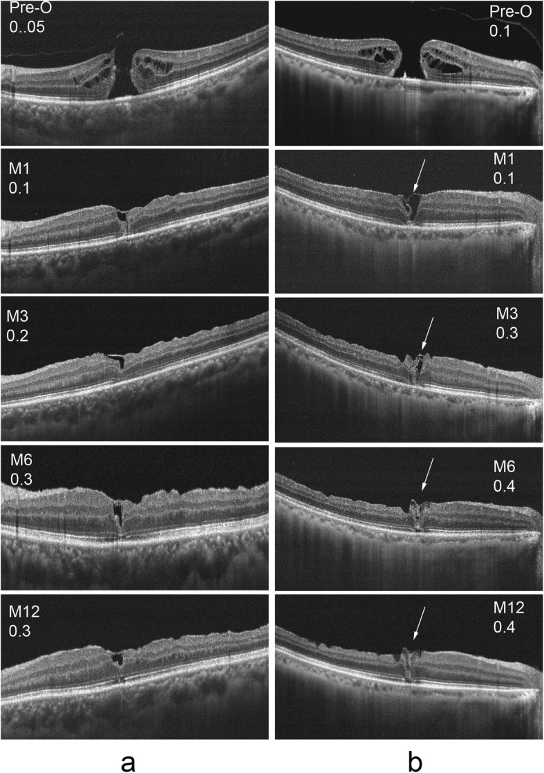
Results: There were statistically significant differences in minimum linear diameter (MLD) of the macular hole and postoperative VA (p = 0.02, 2.81 E-4 respectively) between the two groups. Compared with patients in group A, patients in group B had poorer VA and EZ recovery in the first 12 months after surgery, and a longer ELM recovery period. The OCT results showed that patients in group B had more extensive ILM filling in the macular area after surgery than patients in group A.

Conclusion: The presence of aberrant glial cell proliferation was related to a larger MLD of the IMH, and the filling approach for the ILM during the operation was related to the postoperative healing pattern and vision acuity.

Keywords: Glial cell proliferation; Idiopathic macular hole; Internal limiting membrane flap; Optical coherence tomography; Vitrectomy.


Conflict of interest statement

The authors declare that they have no competing interests.

Figures

Fig. 1
Fig. 1
Examples of the preoperative measurement of the macular hole. This image is a horizontal scan of the OCT results through the macular fovea
Fig. 2
Fig. 2
Preoperative and postoperative visual acuity changes of the two groups at different times
Fig. 3
Fig. 3
The variation trend of the lengths of the ELM (a) and EZ (b) discontinuity in the macular area in the two groups at different times post operation
Fig. 4
Fig. 4
Visual acuity and OCT results pre and post operation of a patient from group A were shown in column a, and the results of a patient from group B were shown in column b. The proliferation of glial cells were shown with arrows in column b
Fig. 5
Fig. 5
Visual acuity and OCT results before and after operation were observed in the two groups. The results of a patient from group A were shown in column a, and the results of three patients from group b were shown in column b, column c and column d, respectively. The patient of column c had a deeper filling depth (shown with arrows) of the ILM during the operation than which of the patient of column d

References

    1. Chang YC, Lin WN, Chen KJ, Wu HJ, Lee CL, Chen CH, Wu KY, Wu WC. Correlation Between the Dynamic Postoperative Visual Outcome and the Restoration of Foveal Microstructures After Macular Hole Surgery. Am J Ophthalmol. 2015;160(1):100–6.e1. doi: 10.1016/j.ajo.2015.03.019. - DOI - PubMed
    1. Kelly NE, Wendel RT. Vitreous surgery for idiopathic macular holes. Results of a pilot study. Arch Ophthalmol. 1991;109:654–659. doi: 10.1001/archopht.1991.01080050068031. - DOI - PubMed
    1. Gu C, Qiu Q. Inverted internal limiting membrane flap technique for large macular holes: a systematic review and single-arm meta-analysis. Graefes Arch Clin Exp Ophthalmol. 2018;256(6):1041–1049. doi: 10.1007/s00417-018-3956-2. - DOI - PubMed
    1. Hayashi H, Kuriyama S. Foveal microstructure in macular holes surgically closed by inverted internal limiting membrane flap technique. Retina. 2014;34(12):2444–2450. doi: 10.1097/IAE.0000000000000252. - DOI - PubMed
    1. Kim Y, Kim ES, Yu SY, Kwak HW. Age-related clinical outcome after macular hole surgery. Retina. 2017;37(1):80–87. doi: 10.1097/IAE.0000000000001162. - DOI - PubMed

LinkOut - more resources