Review
. 2019 Dec 21;14(1):49.
doi: 10.1186/s13024-019-0344-2.

LRRK2 Biology from structure to dysfunction: research progresses, but the themes remain the same

Affiliations
Review

LRRK2 Biology from structure to dysfunction: research progresses, but the themes remain the same

Daniel C Berwick et al. Mol Neurodegener. .

Abstract

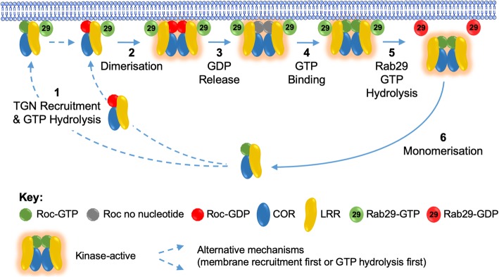
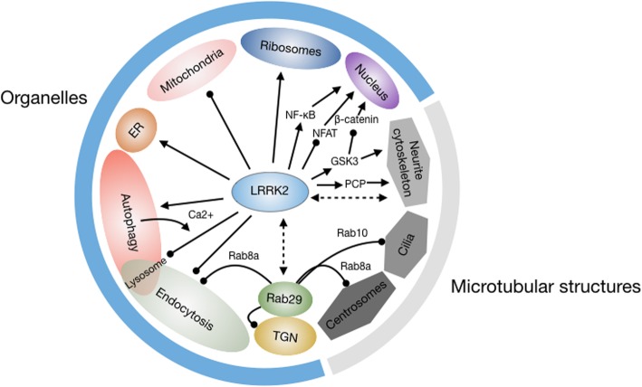
Since the discovery of leucine-rich repeat kinase 2 (LRRK2) as a protein that is likely central to the aetiology of Parkinson's disease, a considerable amount of work has gone into uncovering its basic cellular function. This effort has led to the implication of LRRK2 in a bewildering range of cell biological processes and pathways, and probable roles in a number of seemingly unrelated medical conditions. In this review we summarise current knowledge of the basic biochemistry and cellular function of LRRK2. Topics covered include the identification of phosphorylation substrates of LRRK2 kinase activity, in particular Rab proteins, and advances in understanding the activation of LRRK2 kinase activity via dimerisation and association with membranes, especially via interaction with Rab29. We also discuss biochemical studies that shed light on the complex LRRK2 GTPase activity, evidence of roles for LRRK2 in a range of cell signalling pathways that are likely cell type specific, and studies linking LRRK2 to the cell biology of organelles. The latter includes the involvement of LRRK2 in autophagy, endocytosis, and processes at the trans-Golgi network, the endoplasmic reticulum and also key microtubule-based cellular structures. We further propose a mechanism linking LRRK2 dimerisation, GTPase function and membrane recruitment with LRRK2 kinase activation by Rab29. Together these data paint a picture of a research field that in many ways is moving forward with great momentum, but in other ways has not changed fundamentally. Many key advances have been made, but very often they seem to lead back to the same places.

Keywords: Golgi; LRRK2; Parkinson’s disease; Rab29; Wnt; autophagy; endocytosis; lysosomes; microtubules.


Conflict of interest statement

The authors declare that they have no competing interests

Figures

Fig. 1
Fig. 1
LRRK2 domain structure and function. LRRK2 contains a catalytic core, conferring GTPase activity via the RocCOR domain and kinase activity, embedded in ARM, ANK, LRR and WD40 protein-protein interaction domains. LRRK2 mutations are indicated with a star above the domain structure. LRRK2 is regulated by autophosphorylation of the kinase and Roc domain. Heterophosphorylation by CK1α, IKK and PKA, and dephosphorylation by PP1 regulates interaction with 14-3-3 proteins affecting LRRK2 localisation. Rab29 recruits LRRK2 to the TGN further depicted in detail in Fig. 2.
Fig. 2
Fig. 2
Rab29-dependent LRRK2 activation model. In the presence of GTP-bound Rab29 the equilibrium between monomeric cytosolic LRRK2 and kinase-active dimeric membrane-associated LRRK2 is shifted to the membrane form. 1 Monomeric LRRK2 is recruited to TGN membranes by GTP-bound Rab29. It is not known whether LRRK2 GTP hydrolysis occurs in the cytosol or immediately following membrane recruitment, but the result is an accumulation of monomeric GDP-bound LRRK2 on TGN membranes. 2 The recruitment of LRRK2 to TGN membranes creates a microdomain of high LRRK2 concentration, facilitating LRRK2 dimerisation. 3 While dimerised and kinase-active, LRRK2 releases GDP, 4 GTP exchange occurs, creating dimeric, kinase-active and Rab29-bound LRRK2. 5 Rab29 GTP hydrolysis releases LRRK2 dimers, promoting dissociation from TGN membranes. 6 Decreased LRRK2 concentration in the cytosol favours monomerisation and kinase inactivation. This last step is impaired by pathogenic RocCOR mutations. The representation of LRRK2 as LRR, Roc and COR domains is derived from the LRRK2 GTPase cycle proposed by Deyaert and colleagues, upon which much of this model is built (31).
Fig. 3
Fig. 3
Cell biological processes impacted by LRRK2. As outlined in the main text, LRRK2 has been implicated in regulating processes at membranous organelles and microtubule-based structures, which are shown in the Figure, as are certain LRRK2-mediated signalling pathways that are likely to be involved. Although not mentioned in the main text, the nucleus is included as the subsequent organellar target of transcription factors affected by LRRK2 signalling. The direct interactions between LRRK2 and Rab29, and LRRK2 and microtubules, are depicted with dashed double-headed arrows. Regulatory mechanisms that are clearly inhibitory to the target are depicted with a round-headed arrow; all other relationships (whether activatory, too complex to categorise as inhibitory or activatory, or as yet undefined) are depicted with conventional arrows.

References

    1. Gan-Or Z, Dion PA, Rouleau GA. Genetic perspective on the role of the autophagy-lysosome pathway in Parkinson disease. Autophagy. 2015;11(9):1443–1457. doi: 10.1080/15548627.2015.1067364. - DOI - PMC - PubMed
    1. Elbaz A, Bower JH, Maraganore DM, McDonnell SK, Peterson BJ, Ahlskog JE, et al. Risk tables for parkinsonism and Parkinson's disease. J Clin Epidemiol. 2002;55(1):25–31. doi: 10.1016/S0895-4356(01)00425-5. - DOI - PubMed
    1. Parkinson James. An Essay on the Shaking Palsy. The Journal of Neuropsychiatry and Clinical Neurosciences. 2002;14(2):223–236. doi: 10.1176/jnp.14.2.223. - DOI - PubMed
    1. Zimprich A, Biskup S, Leitner P, Lichtner P, Farrer M, Lincoln S, et al. Mutations in LRRK2 cause autosomal-dominant parkinsonism with pleomorphic pathology. Neuron. 2004;44(4):601–607. doi: 10.1016/j.neuron.2004.11.005. - DOI - PubMed
    1. Paisan-Ruiz C, Jain S, Evans EW, Gilks WP, Simon J, van der Brug M, et al. Cloning of the gene containing mutations that cause PARK8-linked Parkinson's disease. Neuron. 2004;44(4):595–600. doi: 10.1016/j.neuron.2004.10.023. - DOI - PubMed

Publication types

MeSH terms

Substances