. 2019 Dec 21;20(1):86.
doi: 10.1186/s40360-019-0377-8.

SBF-1 inhibits contact hypersensitivity in mice through down-regulation of T-cell-mediated responses

Affiliations

SBF-1 inhibits contact hypersensitivity in mice through down-regulation of T-cell-mediated responses

Wei Chen et al. BMC Pharmacol Toxicol. .

Abstract

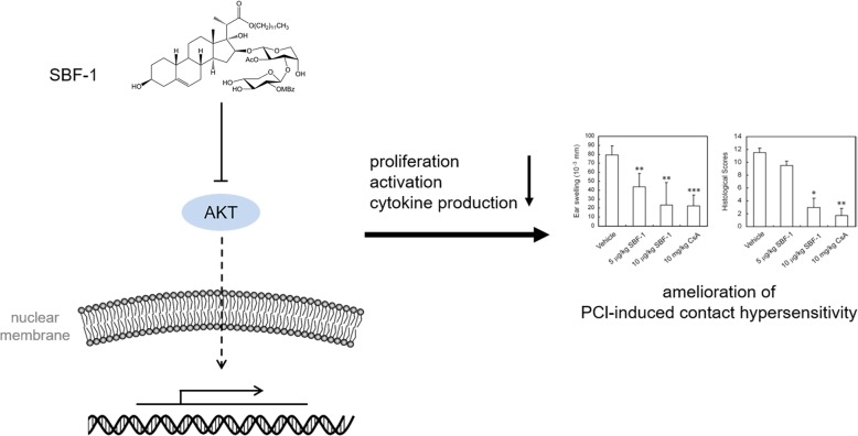
Background: T lymphocytes play an important role in contact hypersensitivity. This study aims to explore the immunosuppressive activity of SBF-1, an analog of saponin OSW-1, against T lymphocytes in vitro and in vivo.

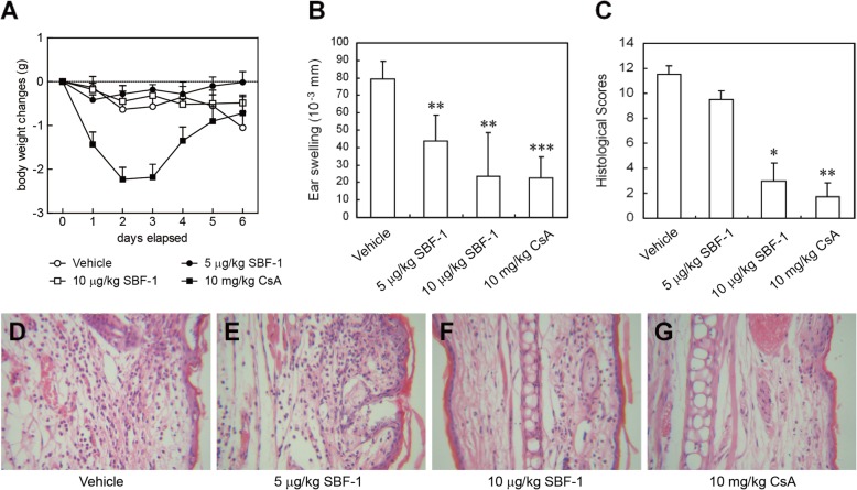
Methods: Proliferation of T lymphocytes from lymph nodes of mice was determined by MTT assay. Flow cytometry analysis was performed to assess T cell activation and apoptosis. Levels of cytokines were determined by PCR and ELISA. BALB/c mice were sensitized and challenged with picryl chloride and thickness of left and right ears were measured.

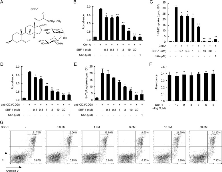
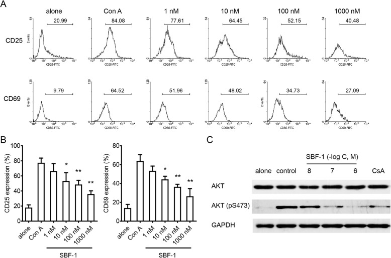
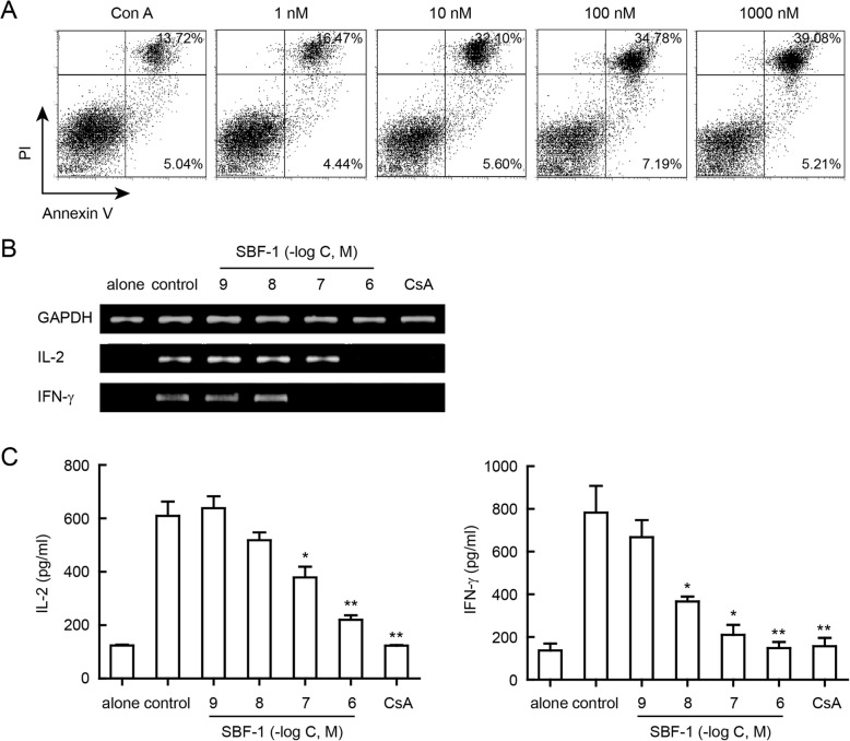
Results: SBF-1 effectively inhibited T lymphocytes proliferation induced by concanavalin A (Con A) or anti-CD3 plus anti-CD28 at a very low dose (10 nM) but exhibited little toxicity in non-activated T lymphocytes at concentrations up to 10 μM. In addition, SBF-1 inhibited the expression of CD25 and CD69, as well as he phosphorylation of AKT in Con A-activated T cells. SBF-1 also induced apoptosis of activated T cells. In addition, SBF-1 also downregulated the induction of the T cell cytokines, IL-2 and IFN-γ in a dose-dependent manner. Furthermore, SBF-1 significantly suppressed ear swelling and inflammation in a mouse model of picryl chloride-induced contact hypersensitivity.

Conclusions: Our findings suggest that SBF-1 has an unique immunosuppressive activity both in vitro and in vivo mainly through inhibiting T cell proliferation and activation. Its mechanism appears to be related to the blockage of AKT signaling pathway.

Keywords: Activated T lymphocytes; Contact hypersensitivity; Immunosuppression; SBF-1.


Conflict of interest statement

The authors declare that they have no competing interests.

Figures

Fig. 1
Fig. 1
SBF-1 markedly inhibited the proliferation of Con A-activated T cells. a The chemical structure of SBF-1. be T cells (3 × 105) were isolated from lymph node of BALB/c mice and then incubated with 0.1–30 nM SBF-1 in the presence of 5 μg/ml Con A bc or 1 μg/ml anti-CD3/CD28 de for 72 h at 37 °C. Cell proliferation was measured at 540 nm by MTT uptake assay b, d or [3H]-thymidine uptake assay (c, e). Data are mean ± SEM of three independent experiments. P values were determined by one-way ANOVA with Tukey’s correction. *P < 0.05, **P < 0.01. fg T cells (5 × 105) were isolated from lymph node of BALB/c mice and then incubated with SBF-1 for 24 h at 37 °C. Cell viability was measured at 540 nm by MTT uptake assay f. Cell apoptosis was analyzed by Annexin V/PI assay staining g. Data are mean ± SEM of three independent experiments
Fig. 2
Fig. 2
SBF-1 suppressed T cell activation through inhibiting AKT signaling. T cells were isolated from lymph node of BALB/c mice and then incubated with SBF-1 in the presence of 5 μg/ml Con A for 24 h at 37 °C. ab Cells were collected and analyzed for CD69 and CD25 expressions by flow cytometry. Data a shown here are one of three different experiments with similar results. Data b are mean ± SEM of three independent experiments. P values were determined by one-way ANOVA with Tukey’s correction. *P < 0.05, **P < 0.01. c Cells were harvested and the whole cell extracts were analyzed by western blotting for AKT and the phosphorylation of AKT. Data shown here are one of three different experiments with similar results
Fig. 3
Fig. 3
SBF-1 induced apoptosis and inhibited T cell cytokines production of activated T lymphocytes. a T cells were isolated from lymph node of BALB/c mice and then activated with 5 μg/ml Con A for 24 h at 37 °C. After being incubated with SBF-1 for 24 h, cells were collected and stained with Annexin V/PI to analyze apoptosis by flow cytometry. Data shown here are one of three different experiments with similar results. bc T cells were isolated from lymph node of BALB/c mice and then incubated with SBF-1 in the presence of 5 μg/ml Con A for indicated time at 37 °C. b T cells were incubated for 24 h, and then they were collected. The mRNA expression of IL-2 and IFN-γ were determined by RT-PCR. Data shown here are one of three different experiments with similar results. c T cells were incubated for 72 h, and then cell culture supernatants were harvested. The levels of IL-2 and IFN-γ were measured by ELISA assay. Data are mean ± SEM of three independent experiments. P values were determined by one-way ANOVA with Tukey’s correction. *P < 0.05, **P < 0.01
Fig. 4
Fig. 4
SBF-1 ameliorated ear swelling in mice with contact hypersensitivity induced by picryl chloride. Contact hypersensitivity was induced in BALB/c mice by PCl. SBF-1 or vehicle control was administered i.p. daily from day 0. a Body weight changes. b Ear swelling. cg Ear tissues harvested from vehicle- or SBF-1-treated mice on day 6 were analyzed for degree of inflammation by H&E (original magnification × 200). c The pathological score. Data are mean ± SEM of six animals per group. P values were determined by one-way ANOVA with Tukey’s correction. *P < 0.05, **P < 0.01. dg Representative photographs of each group: Vehicle d 5 μg/kg SBF-1 e 10 μg/kg SBF-1 f 10 mg/kg cyclosporine A g
Fig. 5
Fig. 5
Schematic representation mechanism of SBF-1’s effects on T cells. SBF-1 suppresses T-cell-mediated immune responses in vitro and in vivo by inhibiting T cell proliferation, activation, cytokine production and inducing apoptosis, which is closely related to its blockage of AKT signaling pathways

References

    1. Bhattacharyya S, Tobacman JK. Arylsulfatase B regulates colonic epithelial cell migration by effects on MMP9 expression and RhoA activation. Clin Exp Metastasis. 2009;26:535–545. doi: 10.1007/s10585-009-9253-z. - DOI - PubMed
    1. Muro R, Nitta T, Kitajima M, Okada T, Suzuki H. Rasal3-mediated T cell survival is essential for inflammatory responses. Biochem Biophys Res Commun. 2018;496:25–30. doi: 10.1016/j.bbrc.2017.12.159. - DOI - PubMed
    1. Smith-Garvin JE, Koretzky GA, Jordan MS. T cell activation. Annu Rev Immunol. 2009;27:591–619. doi: 10.1146/annurev.immunol.021908.132706. - DOI - PMC - PubMed
    1. Speiser DE, Ho PC, Verdeil G. Regulatory circuits of T cell function in cancer. Nat Rev Immunol. 2016;16:599–611. doi: 10.1038/nri.2016.80. - DOI - PubMed
    1. Sell H, Habich C, Eckel J. Adaptive immunity in obesity and insulin resistance. Nat Rev Endocrinol. 2012;8:709–716. doi: 10.1038/nrendo.2012.114. - DOI - PubMed

Publication types

MeSH terms

LinkOut - more resources