. 2019 Dec 6:13:75.
doi: 10.3389/fnsys.2019.00075. eCollection 2019.

Bridging Single Neuron Dynamics to Global Brain States

Affiliations

Bridging Single Neuron Dynamics to Global Brain States

Jennifer S Goldman et al. Front Syst Neurosci. .

Abstract

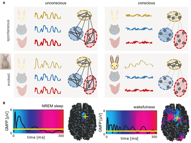
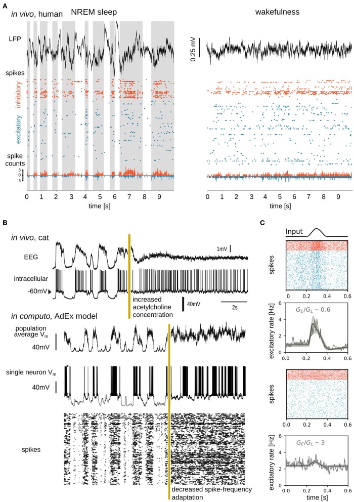
Biological neural networks produce information backgrounds of multi-scale spontaneous activity that become more complex in brain states displaying higher capacities for cognition, for instance, attentive awake versus asleep or anesthetized states. Here, we review brain state-dependent mechanisms spanning ion channel currents (microscale) to the dynamics of brain-wide, distributed, transient functional assemblies (macroscale). Not unlike how microscopic interactions between molecules underlie structures formed in macroscopic states of matter, using statistical physics, the dynamics of microscopic neural phenomena can be linked to macroscopic brain dynamics through mesoscopic scales. Beyond spontaneous dynamics, it is observed that stimuli evoke collapses of complexity, most remarkable over high dimensional, asynchronous, irregular background dynamics during consciousness. In contrast, complexity may not be further collapsed beyond synchrony and regularity characteristic of unconscious spontaneous activity. We propose that increased dimensionality of spontaneous dynamics during conscious states supports responsiveness, enhancing neural networks' emergent capacity to robustly encode information over multiple scales.

Keywords: cerebral cortex; computational neuroscience; coupling; desynchronized; low-dimensional manifold; mean-field models; membrane biophysics; neural network models.


Figures

Figure 1
Figure 1
Complex dynamics associated with conscious brain states provide a potential substrate for neural coding. (A) Schematics of spontaneous (top) and evoked (bottom) dynamics in connected neuronal assemblies encoding different related concepts (different colors) in unconscious (left) and conscious (right) brain states. In unconscious brain states, slow, synchronous, large amplitude oscillations are observed. Stimuli delivered during unconscious states evoke large amplitude, transient responses similar to spontaneous activity. In contrast, during conscious states, asynchronous, irregular firing of neurons results in macroscopically desynchronized, low amplitude signals. Only networks recruited by the perturbation (here, a rabbit) produce lower-dimensional patterns that propagate relatively further in time and space. (B) Global mean-field power (GMFP) recorded with EEG in response to transcranial magnetic stimulation, during deep, non-rapid eye movement (NREM) sleep versus wakefulness. Mean EEG signal is represented by black traces. Background colors represent temporal latency (light blue, 0 ms; red, 300 ms) of maximum current sources, also shown in cortical space on the right, where yellow crosses represent the location of stimulation (right dorsolateral premotor cortex). Reprinted with permission from AAAS (Massimini et al., 2005). If brain dynamics between states may be described in analogy to states of matter, perturbing unconscious brains results in large, brief signals perhaps akin to a small perturbation of a solid, which can displace the solid briefly, but will not modify its internal structure. In contrast, the same perturbation delivered during conscious, liquid-like brain states results in smaller but more complex patterns that propagate further in time and space. Under this interpretation, in coding networks, responses evoked during conscious states could represent a form of transient “crystallization,” consistent with neural trajectories lying on low-dimensional manifolds.
Figure 2
Figure 2
Simple, high-amplitude signals in unconscious brain states are associated with synchronous regular neuronal firing, whereas complex, low-amplitude signals in conscious brain states emerge from asynchronous irregular firing. (A) Data sample from Peyrache et al. (2012), Dehghani et al. (2016), Le Van Quyen et al. (2016), Teleńczuk et al. (2017), and Nghiem et al. (2018b), containing local field potential (LFP; top), spike times (action potentials; middle), and spike counts (bottom) recorded from a human subject during NREM sleep (left) and wakefulness (right). Spikes from inhibitory (orange) and excitatory (blue) neurons were separated and spike counts were calculated in bins of 5ms. Up states shaded in the left panel. (B) Transition between slow-wave (unconscious) and activated (conscious) state dynamics in vivo (top) and in silico (bottom). Experimentally the transition is generated by electrical stimulation of acetylcholine neurons in the pedunculopontine tegmentum (PPT) of anesthetized cat (Volgushev et al., 2011), triggering awake-like, desynchronized dynamics in cortex (Rudolph et al., 2005). A prominent consequence of enhancing cholinergic signaling in cortex is a reduction of spike-frequency adaptation (McCormick, 1992). In silico, a similarly desynchronizing effect can be generated by reducing the parameter responsible for spike-frequency adaptation. Simulated traces shown in the bottom were modified from Destexhe (2009), which used a network of adaptive exponential integrate-and-fire neurons. The average Vm of the network, the Vm of a randomly chosen neuron, and the raster plot of the network are shown. Reproduced with permission from Destexhe (2009). (C) State dependence of network responsiveness. The responsiveness of two spiking networks to a Gaussian pulse is shown. Raster plots display spike times of excitatory (blue) and inhibitory (orange) neurons connected by conductance-based synapses. Population activity (spike counts, thin line), as well as mean-field model (thick lines), and standard deviation (shaded area) of population firing rate generated by a mean-field model developed in di Volo et al. (2019). Responsiveness is found to vary between different network states, obtained by changing the ratio of the time-averaged global excitatory conductance (GE) (Destexhe et al., 2003) to membrane leakage conductance (GL).

References

    1. Arieli A., Sterkin A., Grinvald A., Aertsen A. (1996). Dynamics of ongoing activity: explanation of the large variability in evoked cortical responses. Science 273, 1868–1871. 10.1126/science.273.5283.1868 - DOI - PubMed
    1. Atasoy S., Donnelly I., Pearson J. (2016). Human brain networks function in connectome-specific harmonic waves. Nat. Commun. 7:10340. 10.1038/ncomms10340 - DOI - PMC - PubMed
    1. Boly M., Phillips C., Tshibanda L., Vanhaudenhuyse A., Schabus M., Dang-Vu T. T., et al. (2008). Intrinsic brain activity in altered states of consciousness: how conscious is the default mode of brain function? Ann. N. Y. Acad. Sci. 1129:119 10.1196/annals.1417.015 - DOI - PMC - PubMed
    1. Brookes M. J., Woolrich M., Luckhoo H., Price D., Hale J. R., Stephenson M. C., et al. . (2011). Investigating the electrophysiological basis of resting state networks using magnetoencephalography. Proc. Natl. Acad. Sci. U.S.A. 108, 16783–16788. 10.1073/pnas.1112685108 - DOI - PMC - PubMed
    1. Brown E. N., Lydic R., Schiff N. D. (2010). General anesthesia, sleep, and coma. New Engl. J. Med. 363, 2638–2650. 10.1056/NEJMra0808281 - DOI - PMC - PubMed

LinkOut - more resources