. 2019 Dec 4:10:2767.
doi: 10.3389/fimmu.2019.02767. eCollection 2019.

Relevance of Macrophage Extracellular Traps in C. albicans Killing

Affiliations

Relevance of Macrophage Extracellular Traps in C. albicans Killing

Ana Loureiro et al. Front Immunol. .

Abstract

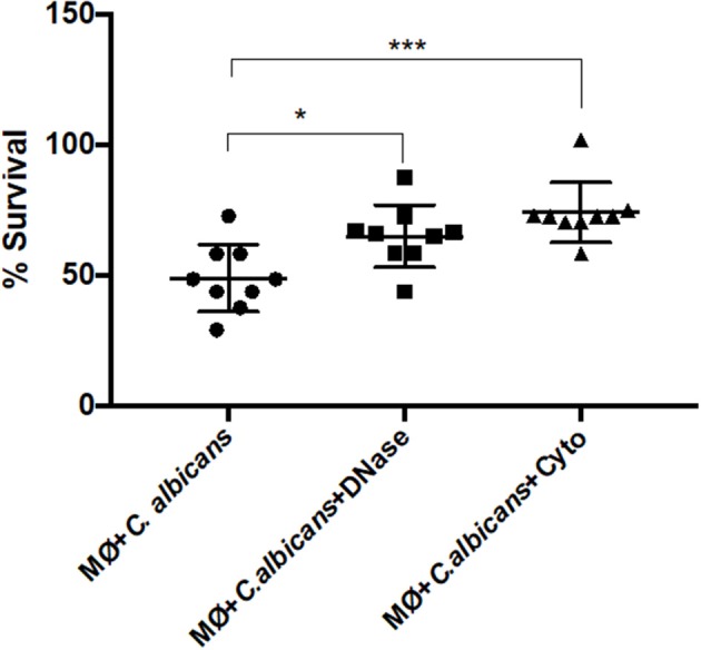
Candida albicans causes systemic life-threatening infections, particularly in immunocompromised individuals, such as patients in intensive care units, patients undergoing chemotherapy, and post-surgical and neutropenic patients. The proliferation of invading Candida cells is mainly limited by the action of the human innate immune system, in which phagocytic cells play a fundamental role. This function is, however, limited in neutropenic patients, who rely mainly on the protective immunity mediated by macrophages. Macrophages have been shown to release extracellular DNA fibers, known as macrophage extracellular traps (METs), which can entrap and kill various microbes by a process called ETosis. In this study, we observed that, upon contact with C. albicans, macrophages became active in phagocyting and engulfing yeast cells. ETosis was induced in 6% of macrophages within the first 30 min of contact, and this percentage increased with the multiplicity of infection until a plateau was reached. After 2.5 h incubation, the presence of extracellular macrophage DNA was observed in approximately half of the cells. This study suggests that the formation of METs occurs before pyroptosis (first 6-8 h) and macrophage cell death (up to 24 h), and thus, METs could be included in models describing C. albicans-macrophage interactions. We also observed that macrophage ETosis and phagocytosis can occur simultaneously and that, in the first hours of infection, both processes are similarly important in controlling the proliferation of yeast cells, this being critical in neutropenic patients. Finally, it can also be concluded that, since C. albicans can degrade DNA, the structural component of METs, yeast extracellular DNase activity can be considered as an important virulence factor.

Keywords: Candida albicans; DNase virulence factor; antifungal activity; macrophage extracellular traps; multiplicity of infection.


Figures

Figure 1
Figure 1
Extracellular fibers observed after macrophages–C. albicans interaction in vitro. Hemacolor staining images of the structures observed following in vitro interaction of C. albicans and J774A.1 macrophage-like cells for 30 min. Macrophages exhibiting typical phagocytic structures (A,B). Extracellular structures resembling macrophage extracellular traps (METs) (C,D). The arrowhead points to extracellular structures that entrap C. albicans cells and the arrow to phagocytosis. Quantification of macrophages exhibiting phagocytosis and METs (E) after infection with C. albicans. *P < 0.05 and ****P < 0.0001 by the Tukey's multiple comparisons test.
Figure 2
Figure 2
DNA release following in vitro incubation of macrophages with C. albicans cells. (1) Fluorescence images of macrophage cells incubated with C. albicans for 30 min and stained with Sytox Green. Control of J774A.1 macrophage-like cells (A) and J774A.1 macrophage-like cells incubated with C. albicans (D). Control cells of primary murine peritoneal macrophages (B) and the same cells incubated with C. albicans (E). Bone-marrow-derived macrophages (BMDM) from BALB/c mice control cells (C) and BMDM infected with C. albicans (F). Quantification of macrophages exhibiting macrophage extracellular traps (METs) (G) after infection with C. albicans. (2) Fluorescence images of J774A.1 macrophage-like cells entrapping C. albicans stained with Sytox Green and anti-H2A-H2B-DNA complex antibody (B); macrophages in more detail entrapping C. albicans yeast cells (blue arrow) and hyphae (white arrow) (C); J774A.1 macrophage-like cells incubated with C. albicans cells and stained only with the secondary antibody and Sytox Green (A). Rotation around the y-axis (a); rotation around the x-axis (b). Red corresponds to the labeling with anti-H2A-H2B-DNA complex antibody; green corresponds to the marking with Sytox Green. (3) Fluorescence images of control J774A.1 macrophage-like cells (A), macrophages infected with C. albicans (B), and macrophages infected with C. albicans and treated with DNase (C) followed by Sytox Green staining. Hemacolor images (D) of macrophages infected with C. albicans and treated with trypsin, with DNase, or not treated. Quantification of macrophages exhibiting METs (E) after infection with C. albicans in the presence and absence of treatments. *P < 0.05 and **P < 0.01 by the Tukey's multiple comparisons test.
Figure 3
Figure 3
Frequency of macrophage extracellular trap (MET) formation with multiplicity of infection. Fluorescence images (arrows point to METs) (A) and quantification of the formation of METs (B) by J774A.1 macrophage-like cells incubated with C. albicans at different MOI (5:1, 10:1, 25:1, 50:1, and 100:1; C. albicans–macrophages) for 30 min and stained with Sytox Green. *P < 0.05, **P < 0.01, and ***P < 0.001 by the Tukey's multiple comparisons test.
Figure 4
Figure 4
Live cell imaging of macrophages incubated with C. albicans. (1A) Representative images of primary murine peritoneal macrophages incubated with C. albicans and Sytox Green, images taken at selected time points during the 2.5-h incubation time (images taken with a 10× objective). (1B) Quantification of Sytox-positive cells over time. Images representative of nine replicates. (2A) Representative images of macrophage cells observed in (1A) where we can discern different MET morphologies. Asterisk represents a diffused extracellular chromatin and number sign a spread extracellular chromatin. (2B) Representative images of the kinetics of MET formation (images from 3). Dashed arrows points to a cell that remained at the puffball-like morphology over the time studied. Solid arrows points to a cell that progressed to comet-like morphology. (3) Representative images of primary murine peritoneal macrophages incubated with C. albicans and Sytox Green, images taken at selected time points during the 4.5-h incubation time (images taken with a 40× objective). Images representative of six replicates.
Figure 5
Figure 5
DNA release in response to non-cellular stimuli and C. albicans. J774A.1 macrophage-like cells stimulated with 200 nM phorbol-12-myristate-13-acetate (PMA), 400 μg/ml N-acetylglucosamine, 200 ng/ml interferon-gamma (IFN-γ), 1,000 μg/ml yeast mannan, 1 μg/ml [lipopolysaccharide 1 (LPS1)], 100 ng/ml (LPS2), or 10 ng/ml (LPS3) LPS, and incubated with C. albicans alone or with yeast and LPS simultaneously. Extracellular DNA was quantified by fluorescence analysis. *P < 0.05 and ****P < 0.0001 by the Tukey's multiple comparisons test, in comparison with macrophages alone.
Figure 6
Figure 6
MET formation by live and heat-killed C. albicans cells. Representative fluorescence images of J774A.1 macrophage-like cells infected with live cells and heat-killed C. albicans (A) and quantification of macrophage extracellular trap (MET) formation (B). Arrows indicate the presence of macrophage extracellular traps (ETs). Evaluation of DNase activity in the supernatants of incubations: agarose gel (C) and intensity quantification (D) of a DNA fragment incubated with supernatant of macrophages alone (1), supernatant of macrophages infected with live C. albicans cells (3), and supernatant of macrophages infected with heat-killed C. albicans cells (5). Negative controls, without DNA addition, of (2) only the supernatant of macrophages, (4) the supernatant of macrophages infected with live C. albicans, and (6) the supernatant of macrophages infected with heat-killed C. albicans. ***P < 0.001 and ****P < 0.0001 by the Tukey's multiple comparisons test.
Figure 7
Figure 7
C. albicans killing by macrophage extracellular traps (METs). C. albicans survival (%) when incubated with J774A.1 macrophage-like cells without any treatment, or with treatment with DNase and/or cytochalasin D. Samples treated with cytochalasin D and DNase I simultaneously were set as the 100% survival control group. *P < 0.05 and ***P < 0.001 by the Tukey's multiple comparisons test.

References

    1. Verma A, Wuthrich M, Deepe G, Klein B. Adaptive immunity to fungi. Cold Spring Harb Perspect Med. (2014) 5:a019612. 10.1101/cshperspect.a019612 - DOI - PMC - PubMed
    1. Antachopoulos C, Roilides E. Cytokines and fungal infections. Br J Haematol. (2005) 129:583–96. 10.1111/j.1365-2141.2005.05498.x - DOI - PubMed
    1. Lorenz MC, Fink GR. Life and death in a macrophage: role of the glyoxylate cycle in virulence. Eukaryot Cell. (2002) 1:657–62. 10.1128/EC.1.5.657-662.2002 - DOI - PMC - PubMed
    1. Qin YL, Zhang LL, Xu Z, Zhang JY, Jiang YY, Cao YB, et al. . Innate immune cell response upon Candida albicans infection. Virulence. (2016) 7:512–26. 10.1080/21505594.2016.1138201 - DOI - PMC - PubMed
    1. Akpogheneta O, Gil-Lamaignere C, Maloukou A, Roilides E. Antifungal activity of human polymorphonuclear and mononuclear phagocytes against non-fumigatus Aspergillus species. Mycoses. (2003) 46:77–83. 10.1046/j.1439-0507.2003.00852.x - DOI - PubMed

Publication types

LinkOut - more resources