Review
. 2019 Nov 18;14(3):164-168.
doi: 10.15420/icr.2019.16.R1. eCollection 2019 Nov.

The Value of Intracoronary Imaging and Coronary Physiology When Treating Calcified Lesions

Affiliations
Review

The Value of Intracoronary Imaging and Coronary Physiology When Treating Calcified Lesions

Yasushi Ueki et al. Interv Cardiol. .

Abstract

Heavily calcified coronary artery lesions hinder the delivery of devices and limit stent expansion, resulting in low procedural success and poor clinical outcomes driven by an increase in restenosis and stent thrombosis. Intracoronary imaging provides a more precise assessment of lesions and is a critical step when deciding whether the lesion needs to be prepared with atherectomy devices. Physiological assessment of lesion significance is an important consideration to avoid unnecessary stenting. This article summarises the current data on the value of intracoronary imaging and functional assessment for coronary calcified lesions and suggests a treatment strategy based on the findings of intracoronary imaging findings.

Keywords: Calcified lesion; fractional flow reserve; intravascular ultrasound; optical coherence tomography; rotablation.


Conflict of interest statement

Disclosure: LR has received research grants from Abbott, Biotronik, HeartFlow, Sanofi and Regeneron, and speaker fees from Abbott, Amgen, AstraZeneca, Bayer, Biotronik, CSL Behring and Sanofi. He is also an unpaid principal investigator of a first in-man study conducted by Biotronik. All other authors have no conflicts of interest to declare.

Figures

Figure 1:
Figure 1:. Images of Calcified Plaque by Intravascular Ultrasound and Optical Coherence Tomography
Figure 2:
Figure 2:. Mechanism of Expansion in a Calcified Lesion
Figure 3:
Figure 3:. Optical Coherence Tomography Findings After Atherectomy Devices
Figure 4:
Figure 4:. A Case with Severely Calcified Lesion Treated by Rotational Atherectomy and Orbital Atherectomy
Figure 5:
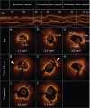
Figure 5:. A Case with Stent Underexpansion Following Rotational Atherectomy
Figure 6:
Figure 6:. Treatment Algorithm for a Calcified Lesion Employing Intracoronary Imaging

References

    1. Wang X, Matsumura M, Mintz GS et al. In vivo calcium detection by comparing optical coherence tomography, intravascular ultrasound, and angiography. JACC Cardiovasc Imaging. 2017;10:869–79. doi: 10.1016/j.jcmg.2017.05.014. - DOI - PubMed
    1. Kini AS, Vengrenyuk Y, Yoshimura T et al. Assessment of fibrous cap thickness by optical coherence tomography in vivo: reproducibility and standardization. J Am Coll Cardiol. 2016;69:644–57. doi: 10.1016/j.jacc.2016.10.028. - DOI - PubMed
    1. Mintz GS, Popma JJ, Pichard AD et al. Patterns of calcification in coronary artery disease. A statistical analysis of intravascular ultrasound and coronary angiography in 1155 lesions. Circulation. 1995;91:1959–65. doi: 10.1161/01.CIR.91.7.1959. - DOI - PubMed
    1. Friedrich GJ, Moes NY, Muhlberger VA et al. Detection of intralesional calcium by intracoronary ultrasound depends on the histologic pattern. Am Heart J. 1994;128:435–41. doi: 10.1016/0002-8703(94)90614-9. - DOI - PubMed
    1. Kostamaa H, Donovan J, Kasaoka S et al. Calcified plaque cross-sectional area in human arteries: correlation between intravascular ultrasound and undecalcified histology. Am Heart J. 1999;137:482–8. doi: 10.1016/S0002-8703(99)70496-5. - DOI - PubMed

LinkOut - more resources