

Semi-quantitative Determination of Protein Expression using Immunohistochemistry Staining and Analysis: An Integrated Protocol
- PMID: 31867411
- PMCID: PMC6924920
- DOI: 10.21769/BioProtoc.3465
Semi-quantitative Determination of Protein Expression using Immunohistochemistry Staining and Analysis: An Integrated Protocol
Abstract
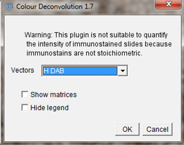
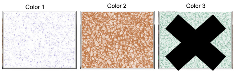
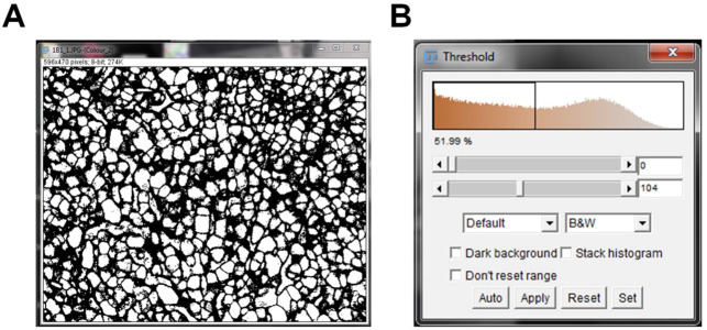
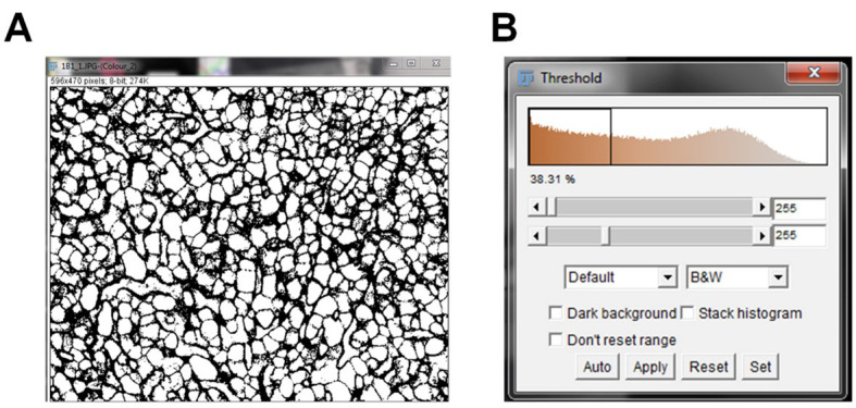
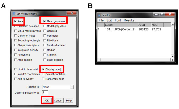
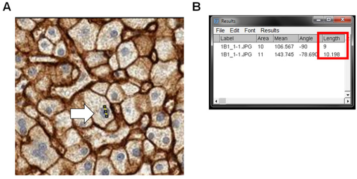
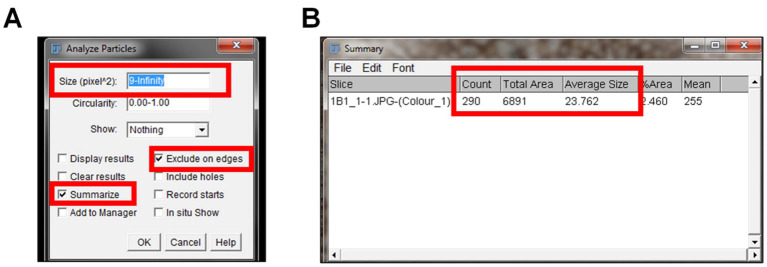
Semi-quantitative IHC is a powerful method for investigating protein expression and localization within tissues. The semi-quantitative immunohistochemistry (IHC) involves using software such as free software ImageJ Fiji to conduct deconvolution and downstream analysis. Currently, there is lack of an integrated protocol that includes a detailed procedure of how to measure or compare protein expression. Publications that use semi-quantification methods to quantify protein expression often don't provide enough details in their methods section, which makes it difficult for the reader to reproduce their data. The current protocol for the first time provides a detailed, step-by-step instruction of conducting semi-quantitative analysis of IHC images using ImageJ Fiji software so that researchers would be able to follow this single protocol to conduct their research. The protocol uses semi-quantitative IHC of organic anion transporting polypeptide (OATP1B1) as an example, and gives detailed steps on how to deconvolute IHC images stained with hematoxylin and 3, 3 - diaminobenzidine (DAB) and how to quantify their expression using ImageJ Fiji. The protocol includes clear steps for a reader so that this method can be applied to many different proteins. We anticipate this method will provide a practical guidance to the reader and make semi-quantification of proteins an easier task to include in publications.
Keywords: DAB staining; Hematoxylin; ImageJ Fiji; Immunohistochemistry; OATP1B1; Semi-Quantification.
Conflict of interest statement
Competing interests No competing financial interests for this study.
Figures
References
-
- Braun M. , Kirsten R. , Rupp N. J. , Moch H. , Fend F. , Wernert N. , Kristiansen G. and Perner S. ( 2013 . ). Quantification of protein expression in cells and cellular subcompartments on immunohistochemical sections using a computer supported image analysis system . Histol Histopathol 28 ( 5 ): 605 - 610 . - PubMed
-
- Chen Y. , Qi Y. , and Xu C. B. ( 2017 . ) A convenient method for quantifying collagen fibers in atherosclerotic lesions by ImageJ software . Int J Clin Exp Med 10 ( 10 ): 14904 - 14910 .
-
- Cregger M. , Berger A. J. and Rimm D. L. ( 2006 . ). Immunohistochemistry and quantitative analysis of protein expression . Arch Pathol Lab Med 130 ( 7 ): 1026 - 1030 . - PubMed
-
- Crowe A. , Zheng W. , Miller J. , Pahwa S. , Alam K. , Fung K. M. , Rubin E. , Yin F. , Ding K. and Yue W. ( 2019 . ). Characterization of plasma membrane localization and phosphorylation status of organic anion transporting polypeptide(OATP) 1B1 c.521 T>C Nonsynonymous single-nucleotide polymorphism . Pharm Res 36 ( 7 ): 101 . - PMC - PubMed
Grants and funding
LinkOut - more resources
Full Text Sources
Other Literature Sources
