. 2019 Dec 3;4(25):21370-21380.
doi: 10.1021/acsomega.9b03035. eCollection 2019 Dec 17.

Antimicrobial Peptide Designing and Optimization Employing Large-Scale Flexibility Analysis of Protein-Peptide Fragments

Affiliations

Antimicrobial Peptide Designing and Optimization Employing Large-Scale Flexibility Analysis of Protein-Peptide Fragments

Neeraj Kumar et al. ACS Omega. .

Abstract

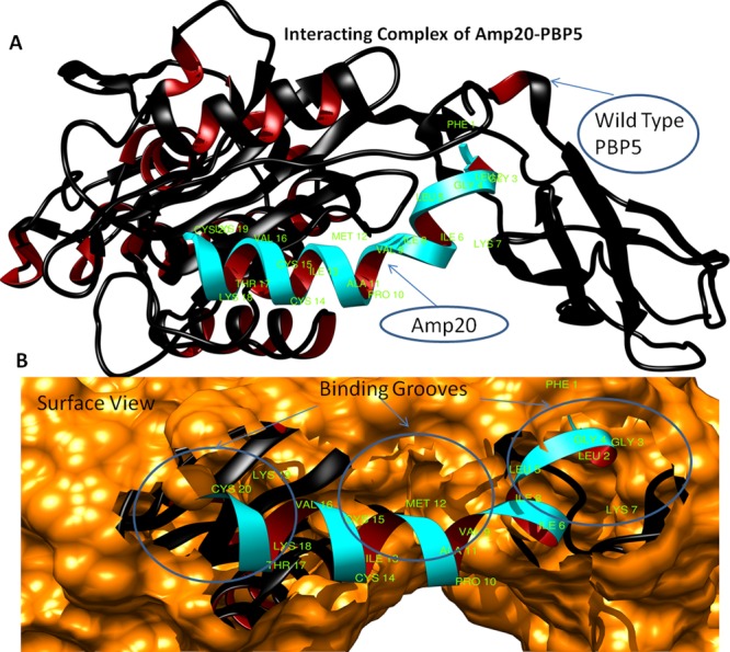
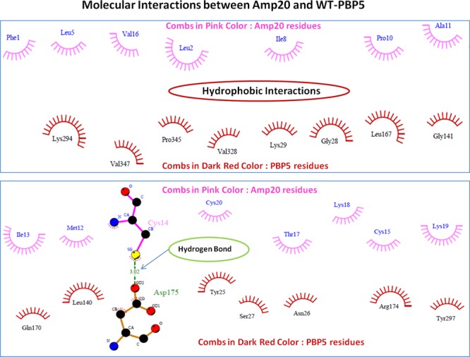
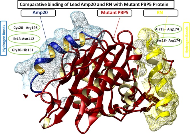
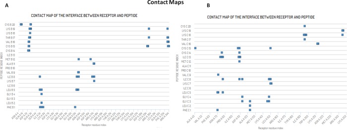
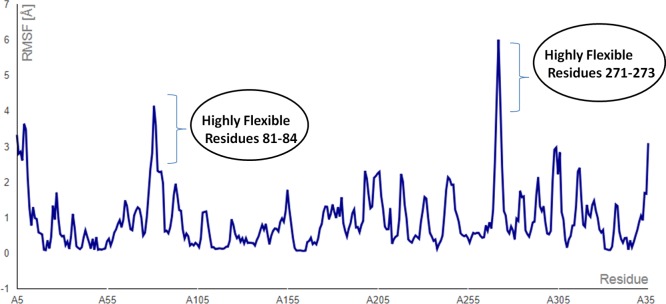
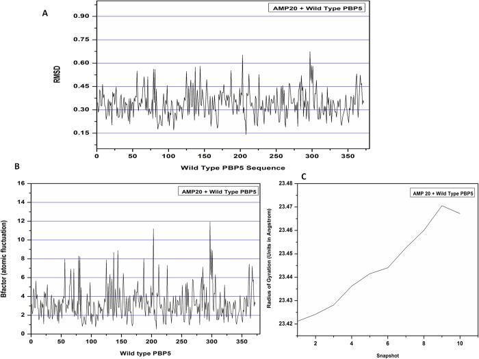
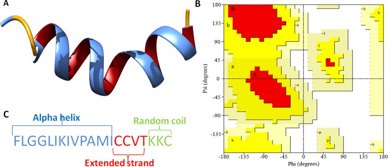
The mankind relies on the use of antibiotics for a healthy life. The epidemic-like emergence of drug-resistant bacterial strains is increasingly becoming one of the leading causes of morbidity and mortality, which gives rise to design a potential antimicrobial peptide (AMP). Here, we have designed the potential AMP using the extensive dynamics simulation since protein-peptide interactions are linked to large conformational changes. Therefore, we have employed the advanced computational avenue CABS molecular docking method that enabled the flexible peptide-protein molecular docking with a large-scale rearrangement of the protein. Lead AMP was investigated against the wild-type (WT) and mutant-PBP5 (MT-PBP5) proteins (antiresistance property). AMP20 showed strong interactions with wtPBP5 and mtPBP5 and involvement of a large number of elements in interactions determined through an atomic model study. Full flexibility analysis showed the stable interaction of AMP20 with both the wild-type and mutant form of PBP5 with root-mean-square deviation (RMSD) values of ∼4.51 and 4.85 Å, respectively. Moreover, peptide dynamics showed involvement of all residues of AMP20 through contact map analysis, and extensive simulation confirmed the stable interaction of AMP20, with lower values of RMSD, radius of gyration, and root-mean-square fluctuation. This study paves the way for a potential approach to design the AMP with amino acid walking and large-scale conformational rearrangements of amino acids.


Conflict of interest statement

The authors declare no competing financial interest.

Figures

Figure 1
Figure 1
(A) Diagram depicting the three-dimensional structure of the PBP5 protein in newcartoon view showing the α-helices in dark blue color with its interior in cyan, pleated sheets in dark red color, and random coils in dark yellow color. (B) Ramachandran plot of the PBP5 protein. (C) Various physiochemical properties of the PBP5 crystal structure (all atoms, solvation energy, torsion, and solvent accessibility) lying in the acceptable region (light blue to blue).
Figure 2
Figure 2
(A) Diagram depicting the accuracy of assessment of the crystal structure of PBP5 by QMEANDisco. QMEAN values (blue region) indicate that the PBP5 model scores higher than experimental structures on average by QMEANDisco. (B) Local quality plot showing the similarity of the native structures and high quality with a score of more than 0.6, expected for high-quality structures. (C) Verify3D web interfaces also confirming the good quality of the structures with minimal deviations in the acceptable range. (D) Structural residues lying under the range of the error-prone or warning region of the plot derived from the Errat protein analysis program.
Figure 3
Figure 3
(A) Structural model of PBP5 with ligand AMP20 (cyan color) interaction and binding sites (green color), newcartoon view. (B) Surface view of binding site pocket residues with best fit confirmation and superimposition of AMP20 (cyan color) to the PBP5 binding grooves.
Figure 4
Figure 4
Depiction of molecular interactions of AMP20 with the PBP5 protein. Involved hydrophobic interactions are shown in the combs in pink color (AMP20) and dark red (PBP5 structure), and hydrogen bonds are shown in green color with a bond length of 3.02 Å.
Figure 5
Figure 5
Comparative binding depiction of AMP20 and ranalexin peptide (RN) at different binding regions of mtPBP5. Notably, AMP20 has strong interactions with three hydrogen bonds, whereas RN binds with two hydrogen bonds.
Figure 6
Figure 6
CABS-dock energy-based top-ranked models obtained for AMP20-PBP5 in the CABS-dock energy graph for top 10 models (shown in different colors). The marker (Model 1) indicates the best model produced in the simulation studies, where the peptide binds close to the PBP5 protein receptor in an open flexible conformation.
Figure 7
Figure 7
Protein-peptide contact maps for the AMP20-PBP5 complex system. (A) Most frequent contacts formed between AMP20 peptide residues with wtPBP5. (B) Contacts formed between AMP20 peptide residues with mtPBP5.
Figure 8
Figure 8
AMP20-PBP5 complex system RMSF (root-mean-square fluctuation) averaged values over the trajectory from the CABS-dock simulation (blue line). Fluctuation run shows the consistent flexible interaction, with a small flexible region at residues 81–84 and 271–273.
Figure 9
Figure 9
Molecular dynamics simulation plot of the AMP20-PBP5 complex. (A) Plot depicting the RMSD structural deviation per residue of the complex. RMSD values show a minimal deviation in the interacting complex. (B) Atomic fluctuation per residue of the target protein with lower deviation. (C) Radius of gyration plot investigation showing the compactness of the protein complex till 10 snapshots. The compactness of protein receptor increased till nine snapshots during the simulation.
Figure 10
Figure 10
(A) 3D crystal structure of the potential antibacterial peptide (AMP20), newcartoon view. (B) Ramachandran plot showing the existence of the AMP20 structure within the favored (94.1% of residues) and allowed (5.9% of residues) regions with no residues in the outlier region. (C) Secondary structure analysis of the peptide.

References

    1. Prestinaci F.; Pezzotti P.; Pantosti A. Antimicrobial resistance: a global multifaceted phenomenon. Pathog. Global Health 2015, 109, 309–318. 10.1179/2047773215Y.0000000030. - DOI - PMC - PubMed
    1. O’Neill J. I. M. Antimicrobial Resistance: Tackling a crisis for the health and wealth of nations. Rev. Antimicrob. Resist. 2014, 1–16.
    1. Iredell J.; Brown J.; Tagg K. Antibiotic resistance in Enterobacteriaceae: mechanisms and clinical implications. BMJ 2016, 352, h6420. 10.1136/bmj.h6420. - DOI - PubMed
    1. Coates A. R. M.; Halls G.; Hu Y. Novel classes of antibiotics or more of the same?. Br. J. Pharmacol. 2011, 163, 184–194. 10.1111/j.1476-5381.2011.01250.x. - DOI - PMC - PubMed
    1. Rasheed M. U.; Thajuddin N.; Ahamed P.; Teklemariam Z.; Jamil K. Antimicrobial drug resistance in strains of Escherichia coli isolated from food sources. Rev. Inst. Med. Trop. Sao Paulo 2014, 56, 341–346. 10.1590/S0036-46652014000400012. - DOI - PMC - PubMed

LinkOut - more resources