. 2020 Oct;16(10):1737-1752.
doi: 10.1080/15548627.2019.1707487. Epub 2019 Dec 27.

BST2 suppresses porcine epidemic diarrhea virus replication by targeting and degrading virus nucleocapsid protein with selective autophagy

Affiliations

BST2 suppresses porcine epidemic diarrhea virus replication by targeting and degrading virus nucleocapsid protein with selective autophagy

Ning Kong et al. Autophagy. 2020 Oct.

Abstract

Interferon-induced BST2 (bone marrow stromal cell antigen 2) inhibits viral replication by tethering enveloped virions to the cell surface to restrict viral release and by inducing the NFKB-dependent antiviral immune response. However, the mechanism by which BST2 uses the selective autophagy pathway to inhibit viral replication is poorly understood. In this study, we showed that BST2 expression was significantly increased during porcine epidemic diarrhea virus (PEDV) infection of Vero cells by IRF1 targeting its promoter. We also showed that BST2 suppressed PEDV replication by binding and degrading the PEDV-encoded nucleocapsid (N) protein. The downregulation of N protein was blocked by macroautophagy/autophagy inhibitors but not a proteasome inhibitor, implying that the N protein was degraded via the selective autophagy pathway. Both the BST2 and N protein interacted with the E3 ubiquitin ligase MARCHF8/MARCH8 and the cargo receptor CALCOCO2/NDP52, and the ubiquitination of N protein was necessary for the degradation of N mediated by the BST2-MARCHF8 axis. The knockdown of MARCHF8 or ATG5 with small interfering RNAs blocked the selective autophagy pathway, rescued the protein abundance of PEDV N in 293T cells, and prevented the inhibition of PEDV replication by BST2 in Vero cells. Together, our data demonstrate the novel mechanism of BST2-mediated virus restriction, in which BST2 recruits MARCHF8 to catalyze the ubiquitination of the PEDV N protein. The ubiquitinated N protein is then recognized by CALCOCO2/NDP52, which delivers it to autolysosome for degradation through the selective autophagy pathway. Abbreviations: 3MA: 3-methyladenine; ATG: autophagy-related; Baf A1: bafilomycin A1; BST2: bone marrow stromal cell antigen 2; CALCOCO2/NDP52: calcium binding and coiled-coil domain 2; CC: coiled-coil; ChIP: chromatin immunoprecipitation; Co-IP: co-immunoprecipitation; CQ: chloroquine; CT: cytoplasmic tail; DAPI: 4',6-diamidino-2-phenylindole; GPI: glycosyl-phosphatidylinositol; hpi: hours post infection; IRF1: interferon regulatory factor 1; ISG: IFN-stimulated gene; MAP1LC3/LC3: microtubule associated protein 1 light chain 3; MARCHF8/MARCH8: membrane-associated ring-CH-type finger 8; MOI: multiplicity of infection; N protein: nucleocapsid protein; PED: porcine epidemic diarrhea; PEDV: porcine epidemic diarrhea virus; RT: room temperature; siRNA: small interfering RNA; STAT: signal transducer and activator of transcription; TCID50: 50% tissue culture infectious doses; TM: transmembrane.

Keywords: BST2; CALCOCO2/NDP52; IRF1; MARCHF8/MARCH8; PEDV; nucleocapsid protein; replication; selective autophagy.


Conflict of interest statement

No potential conflict of interest was reported by the authors.

Figures

Figure 1.
Figure 1.
PEDV infection upregulated BST2 expression in Vero cells and LLC-PK1 cells. (A) Vero cells were infected or mock-infected with PEDV at an MOI of 1 and harvested at the indicated times. The expression of BST2 and PEDV N proteins were analyzed by western blotting. ACTB/β-Actin was used as the sample loading control. (B) BST2 mRNA levels in the same samples (A) were analyzed by real-time PCR. (C) LLC-PK1 cells were treated with universal type I interferon (1,000 U/mL) or infected with PEDV at an MOI of 1 and harvested at the indicated times. The lysates were analyzed by western blotting. (D) BST2 mRNA levels in the same samples (C) were analyzed by real-time PCR. Data are means ± SD of triplicate samples. *p < 0.05, **p < 0.01, ***p < 0.001 (two-tailed Student’s t test).
Figure 2.
Figure 2.
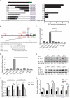
IRF1 directly controls the transcription of BST2. (A) 293T cells were co-transfected with a series of truncated BST2 promoter constructs (−243 to −1, D1 to D8), together with Renilla luciferase reporter vector (pRL-TK-luc), and were analyzed for dual luciferase activity. (B) Regulatory elements in the BST2 promoter region were predicted with the JASPAR vertebrate database (http://jaspar.genereg.net/). Red indicates putative regulatory elements for STAT (signal transducer and activator of transcription), and blue indicates those for IRF (IFN regulatory factor). (C) 293T cells were transfected with BST2 promoter-driven luciferase vector, pRL-TK-luc vector, and plasmids encoding Flag-tagged putative transcription factors (Flag-STAT1, Flag-STAT2, Flag-IRF1, Flag-IRF7, Flag-IRF8, or Flag-IRF9). Samples were collected at 24 h post-transfection and analyzed for dual luciferase activity. (D) 293T cells were transfected with plasmid encoding Flag-STAT1, Flag-STAT2, Flag-IRF1, Flag-IRF7, Flag-IRF8, or Flag-IRF9, followed by real-time PCR to measure the levels of BST2 transcription. (E and F) Vero cells and LLC-PK1 cells were infected or mock-infected with PEDV at an MOI of 0.01, and harvested at the indicated times. The expression of IRF1 was analyzed with western blotting and real-time PCR. ACTB was used as the sample loading control. (G) Vero cells were transfected with IRF1 siRNA or negative control siRNA. The cells were then treated with poly (I:C) (low molecular weight) (10 mg/mL) or PEDV (MOI = 0.01) and the transcription levels of IRF1 and BST2 were analyzed with real-time PCR. Data are means ± SD of triplicate samples. *p < 0.05, **p < 0.01, ***p < 0.001 (two-tailed Student’s t test).
Figure 3.
Figure 3.
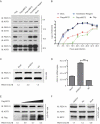
BST2 inhibits PEDV replication in Vero and LLC-PK1 cells. (A) Vero cells were transfected with plasmid encoding Flag-pBST2, Flag-mBST2, or the Flag tag. At 24 h post-transfection, the cells were infected with PEDV at an MOI of 0.01 and harvested at the indicated times. PEDV N protein expression was analyzed with western blotting. ACTB was used as the sample loading control. (B) PEDV titers in the culture supernatants of the Vero cells treated described in (A) were measured as TCID50. (C and D) Vero cells were transfected with mBST2 siRNA or negative control siRNA. At 24 h post-transfection, the cells were infected with PEDV at an MOI of 0.01, and the cell lysates and culture supernatants were then collected for analysis of PEDV N protein expression and viral RNA levels with western blotting and real-time PCR, respectively. (E and F) LLC-PK1 cells were transfected with increasing concentrations of a vector expressing Flag-BST2 (wedge) or pBST2 siRNA. At 24 h post-transfection, the cells were infected with PEDV at an MOI of 1, and the cell lysates were collected for analysis of PEDV N protein expression with western blotting. ACTB was used as the sample loading control. Data are means ± SD of triplicate samples. *p < 0.05, **p < 0.01, ***p < 0.001 (two-tailed Student’s t test).
Figure 4.
Figure 4.
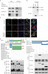
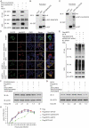
BST2 interacts with PEDV N protein. (A) Vero cells were transfected with plasmids encoding HA-N and Flag-BST2 or empty vectors for 24 h, followed by Co-IP with anti-Flag binding beads and a western blotting analysis with anti-HA, anti-PEDV N, and anti-Flag antibodies. (B) The full length of the BST2 gene and PEDV N gene were cloned into pCold TF plasmid and pCold GST plasmid, respectively. Recombinant proteins were expressed in bacterial strain BL21 (DE3) and purified for the GST pull-down analysis. After adequate washing, proteins eluted from beads were analyzed by western blotting. Input, BST2. (C) 293T cells were transfected with plasmids encoding BST2-GFP and N-mCherry for 24 h. Cell nuclei were labeled with DAPI, and the fluorescent signals were observed with confocal immunofluorescence microscopy. 293T cells were transfected with plasmids encoding BST2-GFP or N-mCherry as control. Scale bars: 100 μm. (D) 293T cells were transfected with plasmids encoding HA-N and Flag-BST2 or the indicated BST2 mutants. They were then analyzed with Co-IP with anti-Flag binding beads and western blotting with anti-HA and anti-Flag antibodies. Throughout was the western blot analysis of whole-cell lysates (WCLs) without immunoprecipitation. ACTB was used as the sample loading control. (E) Co-IP and western blotting analyses of 293T cells transfected with PEDV N or the indicated N mutants, together with a vector encoding Flag-BST2.
Figure 5.
Figure 5.
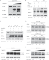
BST2 promotes the autophagic degradation of PEDV N protein. (A) 293T cells were transfected with the vector expressing HA-N and increasing concentrations of a vector expressing Flag-BST2 (wedge) for 24 h. The cell lysates were analyzed with western blotting. ACTB was used as the sample loading control. (B) Vero cells were transfected with HA-N-expressing vector and mBST2 siRNA or negative control siRNA. The cells were then treated with poly(I:C) (low molecular weight) (10 mg/mL) for the indicated times. The cell lysates were analyzed with western blotting. (C) 293T cells were transfected with plasmids encoding Flag-BST2 and HA-N, and the cells were then treated with MG132 (5 mM), bafilomycin A1 (Baf A1; 0.1 mM), chloroquine (CQ; 10 mM), or 3-methyladenine (3MA; 0.5 mM) for 8 h. The cell lysates were then analyzed with western blotting. (D) Vero cells and LLC-PK1 cells were infected or mock-infected with PEDV at an MOI of 1 and harvested at the indicated times. The expression of LC3 was analyzed by western blotting. Vero cells (E) and 293T cells (F) were transfected with the vector expressing HA-N and increasing concentrations of a vector expressing Flag-BST2 (wedge) for 24 h. The cell lysates were analyzed with western blotting.
Figure 6.
Figure 6.
PEDV N protein is degraded by autophagy through the BST2-MARCHF8-CALCOCO2-autophagosome pathway. (A) 293T cells were transfected with plasmids encoding HA-N and MYC-MARCHF8 or MYC-CALCOCO2 for 24 h, and then analyzed with Co-IP with anti-HA binding beads and western blotting with anti-MYC and anti-HA antibodies. Throughout was the western blot analysis of whole-cell lysates (WCLs) without immunoprecipitation. ACTB was used as the sample loading control. (B) The full length of PEDV N gene and MARCHF8 gene were cloned into pCold TF plasmid and pCold GST plasmid, respectively. Recombinant proteins were expressed in bacterial strain BL21 (DE3) and purified for the GST pull-down analysis. After adequate washing, proteins eluted from beads were analyzed by western blotting. Input, PEDV N. (C) The full length of PEDV N gene and CALCOCO2 gene were cloned into pCold TF plasmid and pCold GST plasmid, respectively. Recombinant proteins were expressed in bacterial strain BL21 (DE3) and purified for the GST pull-down analysis. After adequate washing, proteins eluted from beads were analyzed by western blotting. Input, PEDV N. (D) 293T cells were transfected with plasmids encoding N-GFP and MYC-MARCHF8 or MYC-CALCOCO2 for 24 h, and then MYC-MARCHF8 and MYC-CALCOCO2 were labeled with specific primary antibodies and secondary antibodies (red). Cell nuclei were stained with DAPI (blue). The fluorescent signals were observed with confocal immunofluorescence microscopy. 293T cells were transfected with plasmids encoding N-GFP, MARCHF8-MYC, or CALCOCO2-MYC as control. Scale bars: 100 μm. (E) 293T cells were co-transfected with HA-N plasmid, Flag-BST2 plasmid, and Flag-MARCHF8 plasmid or siRNA (hMARCHF8 siRNA or negative control siRNA). Cell lysates were harvested after 3MA (0.5 mM) treatment for 8 h. The proteins were immunoprecipitated with anti-HA antibody, and analyzed with western blotting with anti-ubiquitin (Ub) and anti-HA antibodies. (F) 293T cells were co-transfected with plasmids encoding Flag-BST2 and HA-N and small interfering RNA (BST2 siRNA, MARCHF8 siRNA, ATG5 siRNA, or negative control siRNA), and then analyzed with western blotting with an anti-HA antibody. (G) Vero cells were co-transfected with plasmids encoding Flag-BST2 and small interfering RNA (BST2 siRNA, MARCHF8 siRNA, ATG5 siRNA, or negative control siRNA), and then infected with PEDV (MOI = 0.01) and harvested at the indicated times. Western blotting analysis was performed with a monoclonal antibody against PEDV N protein. (H) PEDV titers in the culture supernatants of the Vero cells treated described in (G) were measured as TCID50.
Figure 7.
Figure 7.
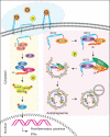
Antiviral mechanism of BST2. (1) BST2 homodimers restrict the release of mature virions from infected cells by positioning one transmembrane domain in the cellular membrane and the other in the virion membrane. (2) YXY motif in the cytoplasmic tail of BST2 interacts with TRAF2/TRAF6. Together, they recruit TAB1, TAB2, and the activated MAP3K7 kinase complex to induce the phosphorylation of CHUK, IKBKB, and IKBKG, thereby activating the canonical NFKB pathway. (3) BST2 recruits the E3 ubiquitin ligase MARCHF8 to catalyze the ubiquitination of PEDV N protein. The cargo receptor CALCOCO2 then recognizes the ubiquitin chains on N protein and delivers the N protein to autolysosome for degradation.

Similar articles

Cited by

References

    1. Chen J, Wang C, Shi H, et al. Molecular epidemiology of porcine epidemic diarrhea virus in China. Arch Virol. 2010;155:1471–1476. - PMC - PubMed
    1. Wood EN.An apparently new syndrome of porcine epidemic diarrhoea. Vet Rec. 1977;100:243–244. - PubMed
    1. Sun RQ, Cai RJ, Chen YQ, et al. Outbreak of porcine epidemic diarrhea in suckling piglets, China. Emerg Infect Dis. 2012;18:161–163. - PMC - PubMed
    1. Li W, Li H, Liu Y, et al. New variants of porcine epidemic diarrhea virus, China, 2011. Emerg Infect Dis. 2012;18:1350–1353. - PMC - PubMed
    1. Debouck P.Pensaert M Experimental infection of pigs with a new porcine enteric coronavirus, CV 777. Am J Vet Res. 1980;41:219–223. - PubMed

Publication types

MeSH terms